
Differential Interference Contrast Image Gallery
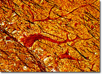
Silver Stained Human Cerebellum
It is often stated that no one ever forgets how to ride a bike, no matter how long it has been since they have actually placed their feet upon the pedals of one. The cerebellum, which stores the trial and error memories obtained while learning a new physical task, is what makes this long-held belief true.
In humans, the cerebellum is a peach-sized, lobed structure located near the base of the brain. It is primarily involved in controlling bodily movement and the development and recollection of physical skills. In order to function properly, the cerebellum must receive information from several parts of the body, such as the eyes, the ears, the limbs, and the cerebrum. The cerebellum, the name of which means “little brain” in Latin, then coordinates all incoming information and makes fine adjustments in motion at the subconscious level. This is how good tennis players can meet the ball in the right place at the right time and return it with appropriate force without having to willfully consider the motions of their muscles.
The two lateral hemispheres of the cerebellum, linked to the larger part of the central nervous system by three wide bands of white matter known as the cerebellar peduncles, are themselves connected by a medial structure called the vermis. Each hemisphere of the cerebellum is further divided into three lobes. The flocculonodular lobe was the first part of the cerebellum to evolve and is primarily associated with maintaining proper balance. The next section to develop was the anterior lobe, which receives sensory input form the spinal cord. The posterior lobe, last to evolve, receives nerve impulses from the cerebrum. Injury to the lobes of the cerebellum or the cerebellar peduncles is generally characterized by muscle weakness, vertigo, ataxia, and tremors.