
Differential Interference Contrast Image Gallery
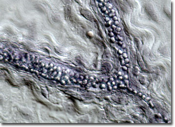
Fat-Stained Adipose Tissue
Adipose tissue is a specialized connective tissue that primarily acts as the main storage site for triglycerides, or fat. In mammals, it exists in two different forms: white adipose tissue and brown adipose tissue.
The body fat that human dieters are generally concerned about is stored in white adipose tissue. Excessive body fat can lead to health problems, but a certain amount of white adipose tissue is necessary for the proper functioning of the body. In addition to storing energy, the tissue, which is usually located directly below the skin, protects the body from impact-related damage to the organs and acts as a heat insulator. In addition to exercise and diet, there are a number of factors that have an apparent effect on the distribution and amount of white adipose tissue stored in a body, including gender, genetics, and race.
Brown adipose tissue derives its name from the characteristic color it gains from densely packed mitochondria and rich vascularization. Although it acts similar to white adipose tissue in some respects, it has the unique ability to generate heat. Consequently, brown adipose tissue is particularly important to small mammals in cold environments and animals in hibernation. The tissue is most prominent, however, in newborns. In fact, though brown adipose tissue comprises about five percent of the weight of human babies, it is virtually nonexistent by the time adulthood is achieved.