|
Darkfield Microscopy Image Gallery
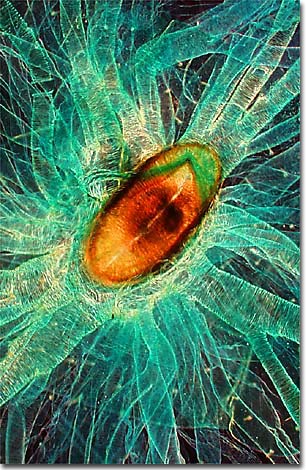
Silkworm Larva Spiracle & Trachea
Stained specimens are often valuable candidates for darkfield microscopy as evidenced by the photomicrograph of a silkworm larva spiracle and trachea, illustrated below.

The specimen is a whole mount, captured with a Nikon Optiphot microscope at 4x under darkfield illumination conditions using a swing-out top lens condenser and a 10 millimeter opaque light stop.
BACK TO THE DARKFIELD GALLERY
Questions or comments? Send us an email.
© 1998-2025 by
Michael W. Davidson and The Florida State University.
All Rights Reserved. No images, graphics, scripts, or applets may be reproduced or used in any manner without permission from the copyright holders. Use of this website means you agree to all of the Legal Terms and Conditions set forth by the owners.
Last modification: Friday, Nov 13, 2015 at 01:19 PM
Access Count Since June 6, 1999: 59006
For more information on microscope manufacturers,
use the buttons below to navigate to their websites:


|

