
Darkfield Microscopy Image Gallery
Liquid Crystalline DNA
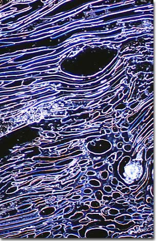
This highly concentrated DNA solution has undergone a series to liquid crystalline phase transitions to form a densely packed hexagonal phase. The photomicrograph was taken with a Nikon Optiphot microscope with a 10x objective in darkfield illumination.
DNA is a very unusual molecule that is shaped like a very long piece of string. The diameter of the DNA molecule is about 25-30 Angstroms (about a billionth of an inch) while the length can exceed a thousand microns (0.025 inch) in some organisms. One of the major problems of molecular biology is how this giant molecule can be packaged into a cell or virus that is far smaller than the length of the DNA molecule. In addition, it is important to the cell to be able to access the DNA molecule for the purposes of genetic control, routine cellular maintenance, and reproduction.
Human DNA is wrapped around a complex of histone proteins, much like beads on a string, in a complex that is termed chromatin. When chromatin condenses for cell division or the production of gametes, chromatin forms higher-order structures that are termed chromosomes. There is a single DNA molecule per chromosome. The DNA of lower organisms such as the virus, bacteria and other prokaryotes, and even some of the more simple eukaryotes does not form highly-organized chromosomes like the ones seen in higher organisms. Lower organisms often package their DNA in reactions that appear to follow the simple laws of thermodynamics: DNA is folded in a compact manner to maximize the entropy of the biological system.
This is often accomplished by highly localized DNA concentrations that drive the molecule into a variety of liquid crystalline states. In the laboratory scientists usually investigate the physical and biological properties of DNA in dilute solution. However, as we have discussed above, DNA exists in vivo in domains where the localized concentrations are very high. As the aqueous solution concentration of DNA is slowly increased, the macromolecule undergoes spontaneous phase transitions to form at least three distinct liquid crystalline phases which are termed lyotropic phase transitions in the liquid crystal field.
The phase of DNA depicted on the banner photomicrograph of this page is a high-density columnar hexatic liquid crystalline phase where the DNA concentration is 400-500 milligrams per milliliter. These are concentrations approaching those found in bacterial nucleoids, dinoflagellate chromosomes, virus capsids, and sperm heads indicating that the DNA in these organelles probably exists in a liquid crystalline state.
Our DNA collection contains over 4000 photomicrographs of liquid crystalline DNA in a variety of phases and concentrations. The various textures exhibited by DNA provide a unique kaleidoscope of patterns that is typical of the way nature is viewed under the microscope.