Darkfield Microscopy Image Gallery
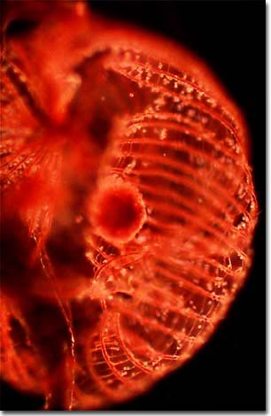
Dictydium cancellatum
The photomicrograph below illustrates the slime mold fungus Dictydium cancellatum under darkfield illumination, imaged with a 10x objective on a Nikon Optiphot microscope.

Mycologists often study slime molds because their fruiting body parts are often similar in appearance to common fungi. These tiny creatures are commonly found in rotted logs and decaying plant matter, where there is an abundant supply of moisture and bacteria. Slime molds produce spores, which germinate to form myxamoebae, a flagellated swarm cell that eventually fuses to generate the plasmodium intermediate and ultimately, the fruiting body.
