Olympus PM-30
Automatic Camera System
The Olympus PM-30 automatic camera system described in this section can easily handle the usual modes of microscopy including brightfield, phase contrast, differential interference contrast, polarized light, Hoffman modulation contrast, and darkfield.

However, the most important facet of this system is the pre-programmed mode featuring two methods for automatically recording fluorescence images, which has classically presented formidable problems for the photomicrographer. The PM-30 camera system can be readily attached to any manufacturer's trinocular observation tube having a vertical phototube with an outside diameter of 25 millimeter (Figure 1). A description of the features included with the PM-30 is outlined in the following paragraphs.
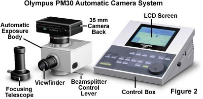
The 35-millimeter film cassette is fitted into the camera back and the protruding strip of film is extended and laid across the film track. When the control unit is activated and the camera back is closed, the film automatically advances to a pre-specified point and is ready for the first exposure. The current exposure number appears in a liquid crystal window on the camera back, facing the user. After each exposure, the film automatically advances to the next frame and is ready for a subsequent exposure. The PM-30 control unit (Figures 2 and 4) offers the option for multiple exposures on the same film frame. When the 24- or 36-exposure film roll is finished, the camera back automatically rewinds the film into the original cassette. After rewinding, small strip of film remains protruding, which is a plus for the do-it-yourself processor or for microscopists who utilize Polaroid instant 35-millimeter film, which must be processed with a special desktop processor. The camera also has an option allowing the user to automatically rewind the film before the roll is completed.
A large-format film adapter is provided and an option for use with 3¼" X 4¼" Polaroid film packs, 4" X 5" single sheet film, or Polaroid 4" X 5" film. The camera automatically senses whether a 35-millimeter or large-format back is attached to the exposure body, and adjusts exposure duration accordingly. If the DX 35-millimeter back and exposure body are being used, the ISO (ASA) speed of the film is automatically set by the control unit, as is the reciprocity factor compensation values. These settings are contained in the memory of the control unit for most current films. Because photomicrographers often prefer to override the film manufacturer's suggested ISO setting or reciprocity factor, the control unit simplifies such resetting. Also, an exposure adjust button can be activated and set for unusual specimen conditions affecting light distribution.
The exposure metering device implanted within the PM-30 body is a 400-element two-dimensional charge-coupled device (CCD) having a spectral sensitivity similar to that of the human eye. The sophisticated electronic circuitry operates the CCD sensor to achieve a high level of sensitivity and accuracy with respect to measurement of illumination intensity at the spectrum of wavelengths encountered during photomicrography.

When the Super Fluorescence mode (SFL) is specified in the control unit, the sensor reads only the areas emitting fluorescing wavelengths. In the regular fluorescence mode (FL), a pre-programmed algorithm functions to reduce the length of exposure, as would be customary for a specimen of varying fluorescence intensities against a dark background of the field of view. The viewfield area being monitored can be set in the control unit to either meter a 30 percent average, a 1 percent spot, or a 0.1 percent spot of a specimen placed in the center of the field. An exposure lock button can be activated, if desired, to allow movement of the metered specimen anywhere in the film frame, while still retaining the selected metered setting.
The control unit has both a visual and a volume-controlled auditory warning to alert the user to pending exposure problems (either overexposure or underexposure). In such instances, exposure is automatically prevented by deactivating the exposure button on the PM-30 control unit. It is also possible to lock in an exposure setting for a series of photomicrographs. The display of the control unit can provide a visual recall reminder of the previous exposure.
The PM-30 exposure body is attached to the vertical tube of a trinocular microscope head, and houses a quiet, vibration-free pulsed electromagnetic shutter. When the expose button is depressed on the control unit, a prism in the exposure body swings completely out of the light path to permit 100 percent of the light reaching the exposure body to reach the film emulsion. Such an arrangement makes shorter exposures possible, which is especially useful in fluorescence microscopy (Figures 2 and 4).

Where desired, the control unit allows the user to program the camera to automatically take a series of three, five, or seven bracketed exposures of various settable time intervals. The photographer can also choose to use a manual setting of time exposure, with a visual countdown of time. In all modes, the exposure time countdown appears on the brightly lighted (brightness and contrast are manually settable) visual display area of the control unit for any exposure slower than one-half second.
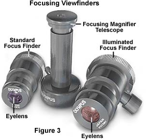
The exposure body accepts either of two diopter-adjustable viewfinders, illustrated in Figure 3. The standard viewfinder has clearly outlined frames that show how much of the field will be included in the photograph. The diopter adjust is rotated by the user to bring the four double crosshairs, centered on the target reticle, into sharp focus. When the specimen and crosshairs are simultaneously in focus, the image will be in focus at the film plane of the camera. The other choice is a brightframed viewfinder, which is especially useful in fluorescence or darkfield photography because the film frame is clearly lighted in either red or yellow. Frame brightness is controllable by means of the job dial on the control unit. A focusing magnifier accessory (Figure 3) overcomes any eye accommodation problems encountered, especially in low-magnification work.
The control unit permits the user to enter and retain, after shutdown, as many as three data settings. With the use of an optional data card inserted into the side of the unit, up to 100 data settings can be stored. An RS232 port allows attachment of the control body to a printer or computer.
Imprinting capability is provided for either the 35-millimeter DX camera back or the large-format attachment, with the controller for entering data already incorporated in the unit itself. For 35 millimeter film, the camera back is replaced with an optional special attachment. There is also a special data adapter is available for large-format imprinting. Eight user-settable alphanumeric characters can be imprinted on the film at any given time.
Because the color temperature setting is so important in color photomicrography, a color temperature metering accessory is provided that attaches to the side of the exposure body. The CTR meter reads the color temperature of the light reaching the exposure body, with the specimen temporarily moved out of the light path. It is then a simple matter for the photomicrographer to set the voltage on the microscope illuminator (some microscopes have automatic voltage setting buttons for this purpose) and to place the appropriate color conversion of color-balancing filters in the light path. This makes certain that the color temperature of the light source is correctly matched to the color film's requirements.

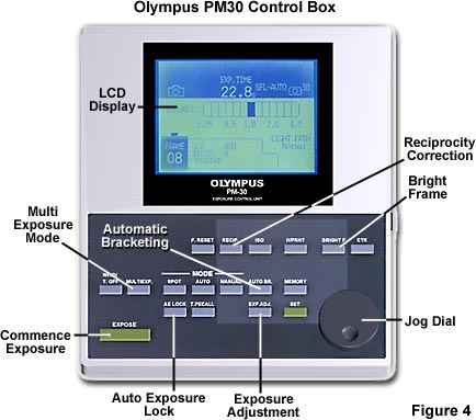
Figure 4 shows the display area and various controls of the PM-30 control unit, which is generally positioned adjacent to the microscope. The unit is a compact 213 X 253 millimeter in size, having a lighted display area that consists of a 92 X 122 millimeter lighted liquid crystal display (LCD) panel. The microprocessor inside the control unit is easily programmed by pressing backlit keys on the unit. The control unit also has tactile sheet controls that are adjustable by means of the large, hand-rotatable jog dial.
The photographer can obtain a wealth of information immediately by simply looking at the lighted, colored display area. This includes the frame number, ASA or ISO setting, reciprocity setting, 30 percent averaging, 1 percent spot, or 0.01 percent spot, light path, exposure in progress, time countdown, exposure adjust setting, and several other variables.
Positioned just below the visual display are the one-touch controls for on-off use: winding, multiple exposure, autoexposure lock, and so forth. On the lighter gray area of the control unit are the button controls, which offer a variety of choices, which can be easily changed by the large rotatable jog dial. These include settings for reciprocity, bracketing exposures, ISO or ASA, and memory. When a button is pressed in the lighter gray area, a display appears. The photomicrographer makes a choice for that item by rotating the jog dial and the set button is then pressed to enter that choice. For example, if the user wishes to change the ISO setting, the ISO button would be pressed. Alternatively, the ISO would by chosen by rotation of the jog dial, and then the set button would be pressed (Figure 4). The PM-30 photomicrographic camera complements the B-Max and older Olympus microscopes, offering a compact system with a host of capabilities.
Contributing Authors
Mortimer Abramowitz - Olympus America, Inc., Two Corporate Center Drive., Melville, New York, 11747.
Michael W. Davidson - National High Magnetic Field Laboratory, 1800 East Paul Dirac Dr., The Florida State University, Tallahassee, Florida, 32310.
