
Bausch & Lomb Fluorescence Microscope
By coupling a laboratory-grade compound microscope with an iron arc lamp and a free-standing filter holder, early twentieth century Bausch & Lomb optical design engineers created a fluorescence microscope that was useful for examining either mineral or organic specimens. Based on a photograph and description in the 1929 Bausch & Lomb microscope catalog, the illustration below was created in 3D Studio Max and Adobe Photoshop.
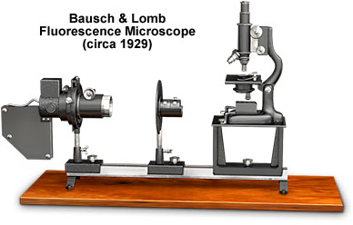
An early example of a fluorescence microscope that is built upon the pioneering work of Köhler, Reichert, and Lehman, the Bausch & Lomb system paved the way for more efficient modern instruments utilizing filter cubes, mercury lamps, and lasers. The Model FFS monocular microscope, a workhorse of the Bausch & Lomb line, features a graceful, long and curved arm, which readily affords manipulation of relatively large specimens. A modified horseshoe base provides stability and is mounted to an adjustable support on the baseboard's V-bar.
Coarse focusing is accomplished by a rack and pinion mechanism that translates the body tube with respect to the rectangular stage, while fine focusing utilizes a patented lever. The body tube, fixed at 160 millimeters, is complemented by a pair of parfocal achromatic objectives (10x and 43x) on a revolving nosepiece. The Bausch & Lomb fluorescence microscope is outfitted with 5x and 10x oculars, and a specialized glass eye cap filter whose purpose is to absorb unwanted background ultraviolet and secondary fluorescence. The eyepiece filter creates a darkfield-like background that enhances specimen fluorescence, but reduces artifacts created by the glass elements.
The substage Abbe condenser lenses are made of quartz, as are the prism reflector and spherical collector lens, because quartz does not absorb appreciable levels of ultraviolet radiation. The condenser is focused by rack and pinion for critical illumination, and the filtered light is directed through a uranium screen that is placed at the iris diaphragm. When the focusing screen fluoresces, the illumination system can be adjusted properly. Before examining specimens the screen is removed. Specimens are mounted on Corex glass slides that pass ultraviolet light without significant autofluorescence. A special filter holder between the iron arc lamp and the quartz prism holds two cells on an adjustable stand. The first cell, which is fitted with quartz windows, is filled with a 25-percent solution of copper sulfate that absorbs long wavelength visible light (red). The second cell, constructed with glass that is transparent to ultraviolet light, absorbs yellow, green, and blue wavelengths and contains a very dilute solution of p-nitrosodimethylaniline, which eliminates the violet wavelengths of light.
BACK TO TWENTIETH CENTURY BAUSCH & LOMB MICROSCOPES
BACK TO TWENTIETH CENTURY MICROSCOPES