
Bausch & Lomb Brinell OM Microscope
A specialized form of the compound microscope, the Bausch & Lomb Brinell OM microscope was designed for measuring the impressions made during the Brinell Ball Test for estimating the hardness of a metal. The 3D Studio Max image presented below is modeled after the original photograph from the review published in the 1929 Bausch & Lomb microscope catalog.
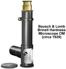
Named after Johann August Brinell, a Swedish engineer who first demonstrated his idea at the Paris Exhibition in 1900, the test provides a hardness number or rating based on pressing a standard size steel ball into a metal specimen using a known force. Unlike more complicated compound microscope models, the Bausch & Lomb Brinell microscope is basically just a body tube mounted on a circular base. A 10-millimeter aperture allows viewing the metal test subject. The tube is fitted with a scale to read 7 millimeters in divisions of a tenth of a millimeter. As an accessory, a 7-millimeter long stage micrometer is provided. The micrometer is etched on opal glass, and is set in a metal disc with rulings in single millimeters, numbered from 0 to 7. Since every tenth line of the ocular micrometer coincides with each millimeter line under the objective, the eyepiece scale is read directly in millimeters and fractions of a millimeter. The stage scale permits comparisons with the eyepiece micrometer and should be checked periodically to determine if recalibration is necessary. The comparison is particularly important after the microscope is used or transported.
The focusing tube slides in the body tube, and carries a Ramsden eyepiece and an achromatic objective, which combine for a total magnification power of 12.7x. Once the user obtains sharp focus, the focusing tube is clamped down with an adjustable ring. Bausch & Lomb shipped the portable Brinell microscope OM with the instrument pre-focused on the viewing field. The interior of the tube surrounding the cutout portion is finished in white to increase the illumination of the specimen, while models OM 17 and OM 18 are designed for occasions when self-contained illumination of the viewfield is required. Model OM 17 is battery-operated, while OM 18 is equipped with cords, connections, and a transformer for 100-volt AC operation. There is also an optional stage micrometer. The draw tube of the Brinell microscope is natural brass, while the outside body tube is smooth black enamel, and the inside is painted white.
BACK TO TWENTIETH CENTURY BAUSCH & LOMB MICROSCOPES
BACK TO TWENTIETH CENTURY MICROSCOPES