
Interactive Java Tutorials
Human Eye Accommodation
Accommodation of the eye refers to the act of physiologically adjusting crystalline lens elements to alter the refractive power and bring objects that are closer to the eye into sharp focus. This tutorial explores changes in the lens structure as objects are relocated with respect to the eye.
To operate the tutorial, translate the Object Position slider and observe how the proximity of the object to the eye changes the shape of the lens. Light rays refracted at the surface of the cornea are further converged by the crystalline lens of the eye. Contraction of the ciliary muscles relaxes the tension on the lens that rounds its shape by virtue of its elasticity while also moving forward slightly. The net effect of the lens changes is to adjust the focal length of the eye to bring the image exactly into focus onto the photosensitive layer of cells residing in the retina. Accommodation relaxes the tension applied through zonule fibers to the crystalline lens, and allows the anterior surface of the lens to increase its curvature. The increased degree of refraction, coupled with a slight forward shift in the position of the lens, brings objects that are closer to the eye into focus.
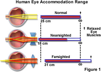
Focus in the eye is controlled by a combination of elements including the iris, lens, cornea, and muscle tissue, which can alter the shape of the lens so the eye can focus on both nearby and distant objects. However, in some instances these muscles do not work properly or the eye is slightly altered in shape, and the focal point does not intersect with the retina (convergent vision). As people age, the lens becomes harder and cannot be properly focused, leading to poor vision. If the point of focus falls short of the retina, the condition is called nearsightedness or myopia, and people with this affliction cannot focus on distant objects. In cases where the focal point is behind the retina, people have trouble focusing on nearby objects, and have a condition called farsightedness or hypermetropia. These malfunctions of the eye can usually be corrected with eyeglasses (Figure 1) using a concave lens to treat myopia and a convex lens to treat hypermetropia.
Convergent vision is not totally physiological and can be influenced by training, if the eyes are not defective. Repetitive procedures can be utilized to develop strong convergent vision. Athletes, such as baseball shortstops, have well-developed convergent vision. In every movement, the two eyes have to translate in unison to preserve binocular vision, with an accurate and responsive neuromuscular apparatus that is not usually subject to fatigue, controlling their motility and coordination. Changes in ocular convergence or head motion are considered in the calculations made by the complex ocular system to produce the proper neural inputs to the eye muscles. An eye movement of 10 degrees may be completed in about 40 milliseconds, with the calculations occurring faster than the eye can reach its intended target. Small eye movements are known as saccades and the larger movements from one point to another are termed versions.
Contributing Authors
Matthew J. Parry-Hill and Michael W. Davidson - National High Magnetic Field Laboratory, 1800 East Paul Dirac Dr., The Florida State University, Tallahassee, Florida, 32310.
BACK TO HUMAN VISION AND COLOR PERCEPTION