
Microscopic Ergonomics Interactive Java Tutorials
Ergonomic Eyepiece Observation Tubes
Eyepiece observation tube height is an important factor that must be seriously considered in the ergonomic design of microscope components and accessories. This tutorial explores the range of currently available ergonomic observation tubes and their extended range of motion, which enables operators of all sizes and heights to comfortably view specimens for lengthy periods of time.
The tutorial initializes with an ergonomically designed binocular observation tube appearing in the window. In order to operate the tutorial, use the Eyepiece Tube Tilt slider to transition the eyepiece tubes through their range of motion with respect to the workstation desk level and microscope stand height. The Eyepiece Tube Length slider enables the eyepiece tubes to be extended or retracted into the main observation head (this slider is not available with all observation tubes contained in the tutorial). A set of radio buttons can be employed to toggle between several ergonomic eyepiece tube designs, including Ergonomic Binocular, Tilting Binocular, Super Widefield, and Stereo Tilting. Each class of observation tube can then be explored with respect to range of motion and ergonomic design.
The major ergonomic factor in the employment of conventional microscopes is that viewing specimens requires users to maintain a flexed neck posture while the hands must remain in a relatively fixed position. From the standpoint of biomechanics, having to maintain even a slight incline of 30 degrees from the vertical can produce significant muscle contractions, muscle fatigue, and pain. Furthermore, it has been documented that pinched nerves can occur when the neck is overextended by this amount. Repetitive motions of the hands and the contact stress of arms resting on a hard surface can cause pain and nerve compression, leading to repetitive stress injuries and/or carpal tunnel syndrome.
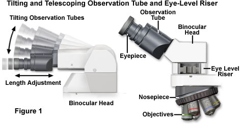
Recent studies have suggested that to permit a more neutral erect working posture, the optical path (distance from the ocular lenses to the specimen being viewed) should lie in the range between 45 and 55 centimeters (18 to 21.5 inches). The eyepieces should be angled no more than 30 degrees above the horizontal plane of the desktop (illustrated in Figure 1). A majority of older microscopes, however, have much shorter optical path length (25 to 30 centimeters or 10 to 12 inches) with the eyepieces angled at 60 degrees above horizontal.
This configuration creates a dilemma for the user. If the microscope is raised to a sufficient height to prevent neck flexion, then the user is forced to bend the wrists into an unnatural position. If the microscope is lowered to bring the stage to a more neutral position, with the forearm parallel to the floor, then the neck is forced to bend. Most workers compensate for this by finding some "happy medium" between the two extreme postures, resulting in discomfort for the neck, shoulders, forearms, wrists, and hands.
Contributing Authors
Robert T. Sutter, Kathleen E. Carr, and Michael W. Davidson - National High Magnetic Field Laboratory, 1800 East Paul Dirac Dr., The Florida State University, Tallahassee, Florida, 32310.
BACK TO ANATOMY OF THE MICROSCOPE