Interactive Tutorials
Channel Photomultipliers
Channel photomultipliers represent a new head-on monolithic design that incorporates a unique detector having a semitransparent photocathode deposited onto the inner surface of the entrance window. The photomultiplier features similar functionality to conventional units, but with dramatically increased sensitivity and high quantum efficiency. Individual photoelectrons released by the photocathode enter a narrow and curved semiconductive channel that serves in place of the traditional dynode chain. Each time an electron impacts an inner wall of the channel, multiple secondary electrons are emitted. This interactive tutorial explores how electrons are multiplied within the conductive chain of a channel photomultiplier.
The tutorial initializes with a cutaway drawing of the channel photomultiplier appearing in the window, and having the photocathode oriented to the left and the anode positioned on the right. Virtual electrons, represented by small yellow spheres, incident on the photocathode are converted into photoelectrons that pass into the channel entrance. In the curved channel, the electrons multiply as they impact the surface of the walls, and eventually are absorbed by the anode. In order to operate the tutorial, use the Gain slider to increase or decrease the number of electrons created each time an electron collides with the channel, and the Tutorial Speed slider to regulate the speed of the electrons passing through the photomultiplier. The tutorial can be halted at any point by clicking on the Stop button, which reverts to a Play button for resumption of the tutorial action.
The spectral response, quantum efficiency, sensitivity, and dark current of a photomultiplier are determined by the elemental composition and geometry of the photocathode. The best photocathodes capable of responding to visible light are less than 40 percent quantum efficient, meaning that at least 60 percent of the photons impacting on the photocathode do not produce a photoelectron and are therefore not detected. Photocathode thickness is an important variable that must be monitored to ensure the proper response from absorbed photons. If the photocathode is too thick, more photons will be absorbed but fewer electrons will be emitted from the back surface. However, if the photocathode is too thin, an abundance of photons may pass through without being absorbed.
The channel photomultiplier addressed in this tutorial is a revolutionary design that eliminates the hundreds of elements necessary to construct a conventional dynode chain. Advantages of this design are lower dark current (picoampere range), increased sensitivity, wider bandwidth, and an extended dynamic range. The monolithic structure provides a high efficiency, self-focusing operation that is also self-biasing. Secondary emission of electrons occurs identically on any portion of the photomultiplier sidewalls, thus optimizing performance.

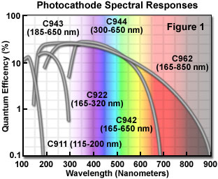
A wide spectrum of photocathode/window combinations are available with the channel photomultiplier to allow use of the device over a wide range of spectral responses (from 115 to 850 nanometers; see Figure 1), using materials that include bialkali, multialkali, and cesium salts (CsI and CsTe, useful only in the ultraviolet region). The most appropriate combinations for confocal microscopy are the bialkali C942 and C943 materials, which operate with high quantum efficiency over the ultraviolet and visible wavelength range from 165 to 650 nanometers. The window materials employed for these photocathodes are either quartz (160-nanometer cutoff) or ultraviolet transparent glass (185-nanometer cutoff). Current amplification by channel photomultipliers with these photocathodes ranges in the neighborhood of 50 to 100 million, and dark current values of approximately 80 picoamperes provide for lower noise levels.

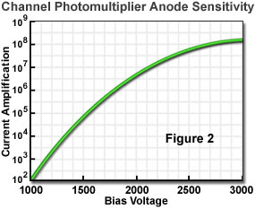
Unlike many older model photomultipliers, the channel photomultiplier requires only a single high voltage supply without the need for an external voltage divider network. At a maximum bias voltage of 3000 volts, anode gain can exceed 100 million (Figure 2), with a sensitivity of 3 million amperes per watt at a wavelength of 410 nanometers using a bialkali photocathode. This performance exceeds that displayed by conventional photomultipliers and avalanche photodiodes by several orders of magnitude. When equipped with specialized photocathode/window combinations, the channel photomultiplier is also a very efficient photon counter. Although most commercial confocal microscopes employ side-on photomultipliers, the channel design may be useful for specialized applications requiring high quantum efficiency and sensitivity.
Contributing Authors
Kenneth R. Spring - Scientific Consultant, Lusby, Maryland, 20657.
Matthew J. Parry-Hill and Michael W. Davidson - National High Magnetic Field Laboratory, 1800 East Paul Dirac Dr., The Florida State University, Tallahassee, Florida, 32310.
BACK TO DIGITAL IMAGING IN OPTICAL MICROSCOPY
