Microscopy Primer
Light and Color
Microscope Basics
Special Techniques
Digital Imaging
Confocal Microscopy
Live-Cell Imaging
Photomicrography
Microscopy Museum
Virtual Microscopy
Fluorescence
Web Resources
License Info
Image Use
Custom Photos
Partners
Site Info
Contact Us
Publications
Home

The Galleries:

Photo Gallery
Silicon Zoo
Pharmaceuticals
Chip Shots
Phytochemicals
DNA Gallery
Microscapes
Vitamins
Amino Acids
Birthstones
Religion Collection
Pesticides
BeerShots
Cocktail Collection
Screen Savers
Win Wallpaper
Mac Wallpaper
Movie Gallery

Overview of Image Processing and Analysis
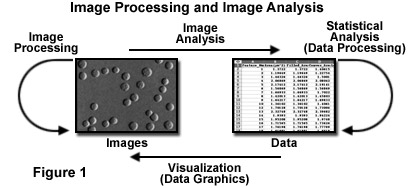
Image Processing operates on images and results in images, with changes intended to improve the visibility of features, or to make the images better for printing or transmission, or to facilitate subsequent analysis. Image Analysis is the process of obtaining numerical data from images. This is usually accomplished by a combination of measurement and processing operations. The data may subsequently be analyzed statistically, or used to generate graphs or other visualizations. Figure 1 summarizes these relationships.

Why do this? There are several reasons; a few are: to assist the human viewer in observing or communicating information in images; to minimize human bias based on wish or expectation; to introduce rigor into the process of obtaining quantitative information as a substitute for anecdote; and not least, to make us better and more aware viewers of images. Unassisted human vision is rarely a reliable scientific tool. Henry David Thoreau said “The question is not what you look at, but what you see.”

The procedures in the following sections are generally applied in the order shown, as appropriate to a given image and final purpose (i.e., skip those steps that are not required in a particular application, but work from the top down). The general sequence of operations is:

  1. Correct or mitigate image imperfections and defects.
  2. Enhance important details by suppressing other information.
  3. Create a binary representation of the structures of interest.
  4. Perform measurements on features and/or overall structure.

Of course, original images should be archived and all processing and measurement steps documented.

Contributing Authors

John C. Russ - Materials Science and Engineering Dept., North Carolina State University, Raleigh, North Carolina, 27695.

Matthew Parry-Hill and Michael W. Davidson - National High Magnetic Field Laboratory, 1800 East Paul Dirac Dr., The Florida State University, Tallahassee, Florida, 32310.


BACK TO INTRODUCTION TO DIGITAL IMAGE PROCESSING AND ANALYSIS

BACK TO MICROSCOPY PRIMER HOME

Questions or comments? Send us an email.
© 1998-2009 by Michael W. Davidson, John Russ, Olympus America Inc., and The Florida State University. All Rights Reserved. No images, graphics, scripts, or applets may be reproduced or used in any manner without permission from the copyright holders. Use of this website means you agree to all of the Legal Terms and Conditions set forth by the owners.
This website is maintained by our
Graphics & Web Programming Team
in collaboration with Optical Microscopy at the
National High Magnetic Field Laboratory.
Last modification: Friday, Nov 13, 2015 at 02:19 PM
Access Count Since July 20, 2006: 20025
For more information on microscope manufacturers,
use the buttons below to navigate to their websites: