Background Subtraction Toolkit Download
The Molecular Expressions digital microscope image Background Subtraction Toolkit is a stand-alone Java application program designed for the Windows operating system, which can be utilized to produce uniform backgrounds for digital images captured with this unique inverted optical microscope.

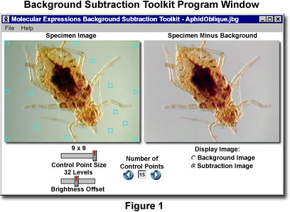
Because of the wide spectrum of illumination modes available in optical microscopy, images can suffer from brightness variations that are manifested by gradients appearing in the background. These fluctuations often lead to contrast and brightness deficiencies in the specimen region and can seriously affect the quality of an otherwise acceptable digital image. The Background Subtraction Toolkit (the user interface is illustrated in Figure 1) is designed to help eliminate background gradients and produce even, uniform backgrounds without affecting the properties of the specimen image. To initiate download of compressed software files, choose a target directory and use the mouse cursor to click on the highlighted links provided below.
Background Subtraction Toolkit Download (File Size: 1,678 Kb) - Recommended for computer display resolutions of 800 x 600 and larger, this Windows software features the essential algorithms to perform background subtraction of digital images having the following file formats: PSD (Photoshop Document), JPG (Joint Photographic Experts Group), BMP (Windows Bitmap), PCX (PC Paintbrush), TGA (Targa Image File), PNG (Portable Network Graphics), and PICT (Macintosh Graphics Files).
Please read our Software License Agreement and disclaimer before downloading and installing any software from this website.
Contributing Authors
Matthew J. Parry-Hill and Michael W. Davidson - National High Magnetic Field Laboratory, 1800 East Paul Dirac Dr., The Florida State University, Tallahassee, Florida, 32310.
BACK TO DIGITAL IMAGING IN OPTICAL MICROSCOPY
