
The Microscope Stand
The microscope stand or frame carries the objectives at the end of the body tube nearest the specimen. Usually there is a nosepiece to accommodate several objectives, each of which can, by means of rotating the nosepiece, be lined up with the body tube opening directly over the specimen in the optical axis of the microscope.
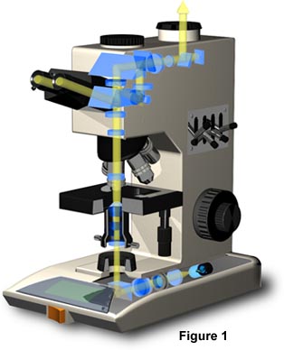
The end of the body tube farthest from the specimen holds the eyepiece. In a straight monocular microscope, there is but one eyepiece at the upper end. In a binocular microscope, by means of prisms, the image projected by the objective is "split" in two and "sent on" to each of the two eyepieces of the binocular. In a trinocular tube, the the observer can, by means of a movable prism, divert the image to the eyes or to the straight tube simultaneously.
In order to minimize vibration, the microscope is constructed with a heavy and rigid base. The microscope tube is attached to the frame. On both sides of the microscope tube or the frame, there are two sets of adjustment knobs; the coarse adjustment knobs for lage increment focusing movements; the fine adjustment knobs for small increment focusing movements. The adjustment knobs serve to bring the objective and the specimen closer together or farther apart. In most microscopes the adjustment knobs raise or lower the stage; in some knobs raise or lower the microscope tube or the nosepiece.
The specimen rests on a stage, rectangular or circle, with an opening in the center to allow light to pass from the lamp through the specimen in order to enter the objective. Often the stage is equiped with a mechanical devicewhich holds the specimen slide in place and can smoothly move the slide back and forth as well as from side to side.
Below the stage is the substage which holds the condenser. The lenses in the condenser serve to concentrate the light from the lamp onto the specimen. The condenser is held in place on a mount which can be moved up and down by means of a focusing knob. The condenser is usually centerable so that all the lenses of the microscope above and below the stage can be kept in alignment. As part of the condenser, or part of the substage, there is an iris diaphragm called aperture diaphragm which can be opened or closed by a knurled ring or lever. The aperture iris diaphragm and the condenser are of critical importance in securing good illumination.
Below the condenser, a mirror is fitted which serves to deflect the light source into the condenser and further through the specimen. The microscope may have a built-in light source and right angle mirror or prism to do this--or the microscope may have a movable mirror which has to be used with an outside light source. The microscope stand has the following functions:
To insure stability and rigidity of the microscope.
To provide the frame for holding the objective and eyepiece at the opposite ends of a body tube.
To make it possible by means of adjustment knobs to focus the microscope objective on the specimen.
To hold the specimen on a stage and enable the specimen to be readily moved on this flat surface.
To carry the substage condenser and mirror which will deflect the light from a lamp up through the specimen.
Contributing Authors
Mortimer Abramowitz - Olympus America, Inc., Two Corporate Center Drive., Melville, New York, 11747.
Michael W. Davidson - National High Magnetic Field Laboratory, 1800 East Paul Dirac Dr., The Florida State University, Tallahassee, Florida, 32310.
BACK TO ANATOMY OF THE MICROSCOPE