
Brightfield Microscopy Digital Image Gallery
Zamia Stem
Zamia is a large genus of cycads, palm-like plants of ancient lineage. The origin of cycads dates back over 200 million years, the trees flourishing on the planet even before dinosaurs first appeared.
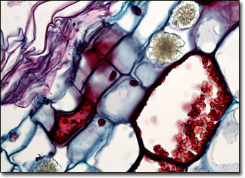
Although ancient cycads were once abundant worldwide, the long-lived and slow-growing modern varieties are significantly diminished in distribution and diversity. These plants, which are now solely found in tropical and subtropical regions, only comprise 250 species in 11 genera, as opposed to the more than 300,000 species of flowering plants that have been identified. Nevertheless, along with ginkgoes and conifers, cycads represent a major order of gymnosperms, or cone-bearing plants. Since cycads are dioecious male and female cones do not appear on the same plant. Instead, the pollen produced by male cones must be carried via the wind or insects to a female cone on another plant in order for fertilization to successfully occur.
Plants of the genus Zamia are primarily found in tropical areas of the Americas, though some also occupy subtropical regions. Indeed, these cycads were once important to many Native American tribes, especially the Seminoles that inhabited parts of Florida. The Seminoles called the plants coonties and often dug up their starchy, turnip-like stems for practical use. After washing them and cutting them into pieces, the stems were typically pounded into a powder. This powder was then repeatedly washed in water so that the starch would separate, resulting in a pasty substance that was fermented and dried back into a powder. The final substance, which today is sometimes referred to as Florida arrowroot, was a diet staple commonly utilized in a manner similar to flour.
BACK TO THE BRIGHTFIELD MICROSCOPY IMAGE GALLERY