Microscopy Primer
Light and Color
Microscope Basics
Special Techniques
Digital Imaging
Confocal Microscopy
Live-Cell Imaging
Photomicrography
Microscopy Museum
Virtual Microscopy
Fluorescence
Web Resources
License Info
Image Use
Custom Photos
Partners
Site Info
Contact Us
Publications
Home

The Galleries:

Photo Gallery
Silicon Zoo
Pharmaceuticals
Chip Shots
Phytochemicals
DNA Gallery
Microscapes
Vitamins
Amino Acids
Birthstones
Religion Collection
Pesticides
BeerShots
Cocktail Collection
Screen Savers
Win Wallpaper
Mac Wallpaper
Movie Gallery

Brightfield Microscopy Digital Image Gallery

Simple Columnar Epithelium

The epithelium is a tissue composed of numerous epithelial cells packed tightly together. These cells, which often come into contact with substances foreign to the body, may vary greatly in shape and function.

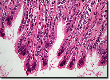
Columnar epithelium can be found along the intestinal tract, spanning from the end of the esophagus to the rectum. This type of tissue also occurs in the ducts of various glands. Epithelial cells are taller than they are wide and contain nuclei along their bases. The membranes that surround them are relatively thin, but can be easily viewed with the aid of a microscope. One of the best-known examples of columnar epithelial cells appears as a covering of the projections called villi found in the small intestine. The functions of these and other cells of the epithelium are various, but essentially involve absorption, secretion, and protection.

BACK TO THE BRIGHTFIELD MICROSCOPY IMAGE GALLERY

Questions or comments? Send us an email.
© 1998-2022 by Michael W. Davidson and The Florida State University. All Rights Reserved. No images, graphics, scripts, or applets may be reproduced or used in any manner without permission from the copyright holders. Use of this website means you agree to all of the Legal Terms and Conditions set forth by the owners.
This website is maintained by our
Graphics & Web Programming Team
in collaboration with Optical Microscopy at the
National High Magnetic Field Laboratory.
Last modification: Friday, Nov 13, 2015 at 02:19 PM
Access Count Since November 25, 2003: 37033
All of the images in this gallery were captured with a QImaging Retiga camera system.
For more information on these cameras, use the button below to access
the QImaging website:
Visit the QImaging website.
For more information on microscope manufacturers,
use the buttons below to navigate to their websites: