
Brightfield Microscopy Digital Image Gallery
Prostate Gland (Younger)
The prostate gland is a vital component of the male reproductive system that surrounds the urethra. Similar in size to a walnut in healthy adults, the organ is actually composed of numerous smaller glands that secrete fluids into sperm stores before they exit the body through the urethra.
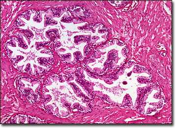
The development of the prostate gland is dependent on the hormones produced by the testes. In infants, the organ is extremely small, approximately the size of a single grain of wheat. During puberty, which usually occurs in males between the ages of 10 and 14, the prostate enters a period of accelerated growth and typically achieves its mature size. However, the prostates of boys who are castrated before this stage in their life never reach their full functioning capabilities or adult dimensions. In unaltered individuals, the prostate continues to change over time and in the fifth decade of life significant enlargement of the gland is common.
Prostatic fluid, which is produced by the prostate gland, is milky and alkaline, containing a number of various constituents, such as zinc, calcium, citrate, phosphate, and enzymes. The substance is believed to be very important to the fertilization process because the other fluids that comprise semen are acidic, and studies have demonstrated that the sperm cells are most active in a relatively alkaline environment. Moreover, the extra medium for the sperm to inhabit protects them and facilitates their greater mobility, making it easier for them to reach and penetrate the egg of a female.
BACK TO THE BRIGHTFIELD MICROSCOPY IMAGE GALLERY