Brightfield Microscopy Digital Image Gallery
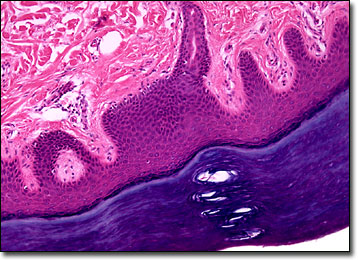
Plantar Skin
Plantar skin is the integument that covers the soles of the feet of humans and other primates. Similar to the skin found along the palms, plantar skin is relatively thick, greatly keratinized, hairless, and filled with a dense collection of sweat glands, which is the reason why the feet and hands are often the first parts of the body to get sweaty when someone is nervous or anxious.

View an image of the plantar skin section at 10x magnification.
Comprised of five morphologically distinct cellular layers, plantar skin is well designed to protect the feet from injury. Indeed, some studies have shown that the skin along the soles of the feet can withstand immensely greater amounts of abrasion or chafing than the skin along most other parts of the body before the pain threshold is reached. Reports also indicate that plantar skin is somewhat resistant to puncture, the integument attempting to shape itself around any sharp objects it comes into contact with. Such characteristics make walking without shoes relatively safe, and in many countries barefoot activity continues to be typical. However, plantar skin is susceptible to bacterial and viral invasions, and a number of problems may occur if infections develop.
Fungi, bacteria, and similar organisms usually thrive in warm, moist environments, and places such as gyms and public showers are where most people acquire infections in the skin that lines the soles of the feet. One of the most familiar infections that affect the feet is athlete’s foot, a condition caused by a fungus and characterized by redness, scaling, and itching of the plantar skin and in the areas between the toes. Athlete’s foot, also known as tinea pedis, may often be cured or improved through repeated application of over-the-counter anti-fungal creams and powders. Plantar warts, however, which are another common type of plantar skin problem, are caused by a virus and often must be removed via more serious methods, such as surgery, cryotherapy, acid application, or laser treatment.
BACK TO THE BRIGHTFIELD MICROSCOPY IMAGE GALLERY
