Brightfield Microscopy Digital Image Gallery
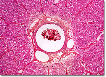
Optic Nerve
Human vision is a very complex process that involves the nearly simultaneous interaction of the eyes and the brain through a network of neurons, receptors, and other specialized cells. Vital to this interactive communication are the optic nerves, which carry nerve impulses from the retinas to the thalamus and other parts of the brain.

The optic nerve, which arises from the junction of visual nerve fibers in the optic disk located in the back of the eyeball, is the second of twelve distinct paired cranial nerves. In this area there is a lack of receptor cells, resulting in what is commonly called the blind spot of the eye. From this exit point, the optic nerve extends to the optic chiasma, located on the underside of the hypothalamus. It is at this X-shaped structure that the optic nerve from each eye meets, part of the fibers of each crossing over to the other side of the brain and the other half maintaining their original orientation. Subsequent to their partial convergence, the nerve fibers of the optic nerves pass on to the visual cortices, where the processing of visual information continues.
Each optic nerve contains approximately one million nerve fibers, a number substantially lower than the more than one hundred million receptors that are located in the retina. Thus, it is generally assumed that a significant amount of information processing takes place within the retina, greatly decreasing the number of signals that must be transferred to the brain via the optic nerve. Yet, any damage to the optic nerve may be as detrimental to one’s sight as an injury to the retina. The extent to which loss of sight occurs in such instance does, however, depend upon the severity of the damage, as well as the precise location in which it occurs.
BACK TO THE BRIGHTFIELD MICROSCOPY IMAGE GALLERY
