
Brightfield Microscopy Digital Image Gallery
Mammalian Testes
The testes are important male reproductive glands that are generally located within the body of vertebrates. Sexually mature mammals typically possess a special sac called the scrotum which houses the testes, and a few varieties of the animals are capable of retracting the glands at will.
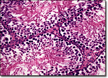
The human testes are oval-shaped organs that weigh about 25 grams each. Though only about two inches long and one inch in diameter, each of these reproductive glands is comprised of approximately 800 seminiferous tubules. In younger individuals, the tubules typically exhibit a simple shape, but after sexual maturity is reached, the structures become branched and coiled. The fully developed tubules are capable of generating sperm through the stimulation of the spermatogonia, or sperm-producing cells. The epididymis is the sexual structure where all of the tubules located in each testis meet. This tube-shaped organ extends into the ductus deferens, which is the primary target for most medical male sterilization procedures.
In addition to spermatogonia, a number of other cells are associated with the testes. Sertoli cells, for instance, are found in both the testes of young boys and adult males. These specialized cells are responsible for supporting and protecting the spermatogonia. Also Leydig cells, which comprise the interstitial tissue between the seminiferous tubules, are believed to secrete androgens that are important for the maturation of the male reproductive apparatus as well as the development of secondary sex characteristics. The number and physical characteristics of Leydig cells vary to a significant extent in different species, but are always influenced by the action of the pituitary gland.
BACK TO THE BRIGHTFIELD MICROSCOPY IMAGE GALLERY