
Brightfield Microscopy Digital Image Gallery
Mammalian Smooth Muscle Tissue
The muscle tissue in mammals and other higher animals is usually described as either striated, cardiac, or smooth depending on its function and appearance. The most studied of these types of tissue is striated muscle, but a significant body of information has been compiled on smooth and cardiac muscles as well.
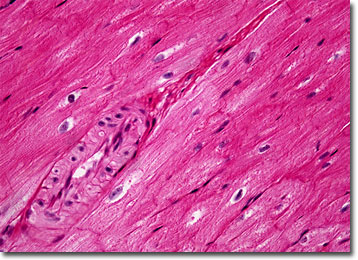
Smooth muscle is typically comprised of numerous elongate spindle-shaped cells, each of which contains a single nucleus located in its center. As indicated by its name, the tissue displays no striations or other distinct patterns under the microscope. The contraction of smooth muscle is slow and generally under the control of the autonomic nervous system, resulting in its alternate moniker, involuntary muscle. Most of the body’s hollow internal organs, including all of the parts of the digestive system, are lined with smooth muscle.
More varied than other types of muscle tissue, smooth muscle may exhibit a number of different characteristics. In addition to their spindle-like form, for instance, the cells that comprise smooth muscle may also be shaped like ribbons or rods. The action of the cells also varies somewhat. For example, the smooth muscles located in the intestines work in conjunction with one another and may stimulate their own contraction, while in other areas the cells may be controlled individually by the body’s nervous system.
BACK TO THE BRIGHTFIELD MICROSCOPY IMAGE GALLERY