
Brightfield Microscopy Digital Image Gallery
Mammalian Graafian Follicle
Graafian follicles are the rounded vesicles found in the cortex of the mammalian ovary. Sometimes alternatively referred to as ovarian follicles, these important reproductive structures enclose and protect developing ova.
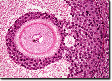
During the youth of a female, several hundred thousand graafian follicles may be present in the ovaries, each of which contains an oocyte surrounded by a single stratum of cells. By the time puberty is reached, however, most of these follicles have deteriorated and collapsed. Those that remain develop to maturity one at a time in a monthly cyclical process. A substance called the follicle-stimulating hormone that is emitted by the pituitary gland is the factor that facilitates the beginning of this process, ensuring the follicles growth and maintenance. The luteinizing hormone, which is also secreted by the pituitary gland, is also involved in the maturation of the graafian follicle, eventually stimulating the secretion of estrogens by the follicle and causing the vesicle to burst.
When a graafian follicle bursts, the oocyte it contains is released into the peritoneal cavity, from which it travels to the uterine tube. This event occurs about halfway through the human female reproductive cycle and is commonly known as ovulation. Subsequent to ovulation, the ruptured graafian follicle is quickly transformed into a vascularized body called the corpus luteum. This structure, which is chiefly influenced by the luteinizing hormone, generates progesterone and estrogens until it begins to degenerate at the end of the reproductive cycle if fertilization does not transpire.
BACK TO THE BRIGHTFIELD MICROSCOPY IMAGE GALLERY