
Brightfield Microscopy Digital Image Gallery
Mammalian Cerebellum
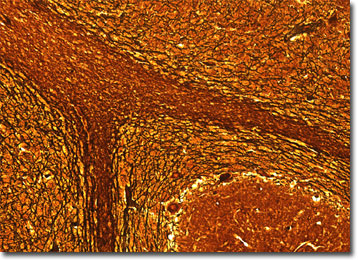
The cerebellum is part of the hindbrain located behind the pons. The lobed structure, which in humans is about the size of an adult fist, functions chiefly in the control of bodily movement and the development and recollection of physical skills, such as riding a bike or swimming.
The two lateral hemispheres of the cerebellum are each composed of three different lobes that evolved at different times. The earliest to develop was the flocculonodular lobe, which is intricately involved in the maintenance of balance. The next region of the cerebellum to evolve was the anterior lobe, a structure that is responsible for receiving sensory information from the spinal cord. The posterior lobe, which accepts nerve impulses from the region of the brain called the cerebrum, was the last part of the cerebrum to evolve. Together these lobes are connected to the rest of the central nervous system by the cerebellar peduncles, three broad bands of white matter.
Fast, effective communication between the cerebellum and other parts of the brain and body is essential for smooth, coordinated movements to take place. When a baseball player swings a bat, for instance, his eyes must judge how fast the ball is approaching, his brain must send the intent of movement to the muscles, and the cerebellum must coordinate all incoming information in such a way that the arms move around the body at the appropriate level and with sufficient force to hit the ball. In order to do this, the cerebellum must make corrections almost instantaneously depending on the information it receives, enabling good players to hit a curve ball even if they were expecting a different type of pitch.
BACK TO THE BRIGHTFIELD MICROSCOPY IMAGE GALLERY