
Brightfield Microscopy Digital Image Gallery
Hemlock Leaf
There have been about 14 species of hemlock trees identified and classified, all of which are members of the genus Tsuga of the family Pinaceae. A variety of other organisms, such as poison hemlock and water hemlock, are commonly referred to by the same name, but are members of the plant family Apiaceae.
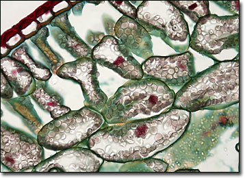
True hemlocks are tall, coniferous trees that exhibit short, blunted leaves and slender branches. The branches, which may be horizontal to the ground or drooping, are generally covered in reddish to purplish brown bark and are tipped with small, egg-shaped cones that hang downwards. Native to North America and Asia, some hemlock species have been greatly reduced in number due to human development. For instance, the common hemlock of eastern North America, scientifically described as T. canadensis, has been commercially valuable as a source of tannin and a type of tea may be made from its young needles, but the tree is not as prevalent as it once was.
The hemlock tree typically found in western North America is T. heterophylla, also known as Prince Albert’s fir and hemlock fir. Trees of this species are the tallest in the genus, often growing as many as 200 feet high. Lumber derived from them is superior to that of other hemlocks and is comparable in quality to pine. The wood of the western hemlock was formerly utilized heavily to construct sugar and flour barrels since it is free from resinous materials, but is used more often today for paneling, boxes, crates, and general construction.
BACK TO THE BRIGHTFIELD MICROSCOPY IMAGE GALLERY