Brightfield Microscopy Digital Image Gallery
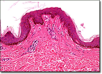
Heavily Pigmented Skin
Almost all living organisms exhibit pigmentation, and some are capable of adapting the characteristic based on their environment. Both flora and fauna are capable of synthesizing pigments, and animals have the additional capability of deriving pigments from their diet.

View an image of the heavily pigmented skin section at 10x magnification.
Pigments are what dictate the coloration of an organism. The chief pigments found in animals include guanine, which is expressed as the color white or as an iridescent hue, the carotenes, which range from yellow to red, the melanins, which are perceived as brown or black, and the hemes, which are responsible for the reddish hue of blood hemoglobin. Some of these pigments function in various physiological activities as well as providing color. Most notable in this regard are the carotenes, which are important in the vitamin synthesis of animals, as well as chlorophyll synthesis in plants.
In humans, melanins are largely responsible for the relative darkness of the skin, as well as the hair and the irises of the eyes. High levels of localized melanin are also the cause of freckles and moles. The amount of melanin pigments in the body are usually a function of genetics, but may be altered by various means. A suntan, for instance, is an indication of the overproduction of melanin caused by exposure to ultraviolet light. Certain disorders, such as Recklinghausen’s neurofibromatosis, which involves the improper storage of melanin in melanocytes, can also cause unusually high levels of pigmentation. Underproduction of melanin or a related absence of melanocytes may result from a number of conditions as well, including albinism, vitiligo, and phenylketonuria.
BACK TO THE BRIGHTFIELD MICROSCOPY IMAGE GALLERY
