
Brightfield Microscopy Digital Image Gallery
Frog Striated Muscle Tissue
Of the three different muscle types found in frogs and most other higher animals, the best understood is the striated muscle. Also known as voluntary, striped, and skeletal muscle, this tissue type is responsible for the movement of an animal’s skeletal structure.
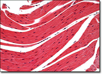
Striated muscle is the most prevalent muscle type within the body, comprising a large proportion of the total body weight of frogs. This type of muscle is comprised of numerous long, cylinder-shaped fibers that exhibit a significant level of organization. Each fiber exhibits alternating dark and bright bands, or striations, that can be clearly seen with the aid of a microscope and multiple nuclei. Each fiber is also encircled by a multilayered structure known as the sarcolemma and contains sarcoplasm, a type of cytoplasm rich in adenosine triphosphate (ATP), phosphagens, enzymes, and other important molecules.
Attached to the body’s skeleton via flexible tendons, striated muscles are usually arranged in pairs. Each member of a pair of pulls in the direction opposite of the other one, providing precise control of the skeletal structure. For instance, the biceps are responsible for flexing the forearm and the triceps are responsible for extending the same part of the body. The contraction of striated muscle is stimulated by electrical impulses sent out by the nervous system and requires energy from ATP. Some of the bodily sources of this important substance include glycolysis, oxidative phosphorylation, and the Krebs cycle.
BACK TO THE BRIGHTFIELD MICROSCOPY IMAGE GALLERY