
Brightfield Microscopy Digital Image Gallery
Fern Spores
Ferns are primitive, spore-producing vascular plants with true roots, stems, and complex leaves. Though they were the dominant type of vegetation found on Earth during the Carboniferous period, ferns are somewhat decreased in distribution in modern times, but are still found around the world.
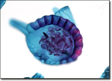
The reproduction of most ferns involves an alternation of sexual and asexual generations. Few people can recognize the fern, however, in its sexual form, which appears as a tiny kidney-shaped plant referred to as the gametophyte or prothallium. The asexual form, known as a sporophyte, is representative of the fern as it is most commonly known. Sporophyte ferns are capable of reproducing both by vegetative cloning and by the production and dispersion of spores, which usually form on the underside of the leaves in clusters of spore cases called sporangia or sori. Depending on species, sori may appear on all or only a fraction of the plant’s leaves. When these structures dry out, they rupture, releasing the numerous spores they contain into the air where they may be carried by the wind to new locales for germination.
Fern spores are typically covered in a thick cell wall, but may exhibit a wide variety of characteristics, many of which play a critical role in determining the taxonomic classification of species. The basic shape of these reproductive cells may, for instance, be either tetrahedral or bilateral, while their walls may be either smooth or variously patterned. The reason for the diversity of spores in such regards is not fully understood by modern scientists, many of the differences that occur among the cells seeming to have no functional purpose.
BACK TO THE BRIGHTFIELD MICROSCOPY IMAGE GALLERY