
Brightfield Microscopy Digital Image Gallery
Epididymis
The epididymis is an elongated paired structure that is part of the male reproductive system. Located along the back of the testes, the epididymis is responsible for transporting the sperm produced by the reproductive glands.
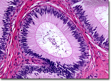
Crescent-shaped, the epididymis is part of what is generally referred to as the sperm canal. This canal, which also comprises the ductus deferens and the ejaculatory ducts, spans the area between each testis and the urethra. A number of small ducts, or ductules, within the canal are responsible for transporting spermatozoa through the body before it is ejaculated. A muscular sheath that surrounds each ductile as well as ciliated columnar cells help facilitate this process.
The epididymis, which receives a constant supply of blood from a branch of the testicular artery and is divided into three basic regions, is believed to function in multiple ways. The largest part of the epididymis, known as the head, is located atop the testis, while the smallest part, commonly called the tail, is located where the epididymis separates from the gland. Of intermediate size is the body, which is attached to the rear of the testis and spans its length. The head and body of the epididymis are essential for the maturation of sperm and the tail serves as a storage site for the reproductive cells. The various parts of the epididymis may also be involved in the removal of excess fluid surrounding sperm, but the evidence for this function of the reproductive structure is not conclusive.
BACK TO THE BRIGHTFIELD MICROSCOPY IMAGE GALLERY