
Brightfield Microscopy Digital Image Gallery
Cerebrum
Humans have been intrigued by the origin of intelligence for thousands of years, although some early philosophers believed that the acumen was located in the heart, not the brain. Significant insight into the true function of the brain was not gained until the eighteenth century when Italian physiologist Luigi Galvani demonstrated that electricity existed inside brain cells.
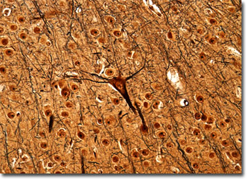
View an image of the cerebrum section at 20x magnification.
The many complex functions carried out by humans on a daily basis are now understood to be regulated by the brain, which has changed significantly over the course of evolution. Today, the vast majority of the brain�s weight (about 85 percent) is dedicated to the cerebrum. In early man, however, the cerebrum was not as well developed, consisting of only about a third of its current weight. This significant physical change has been accompanied by important changes in the mental processes, humans becoming increasingly capable of more and more complex activities over time. Indeed, most scientists believe it is the relative size of the cerebrum that most differentiates humans from other animals, since it is considered the origin of all conscious activity. It is important to note, however, that the purely physical size of the brain is not an indication of intelligence, since, for example, the brain of an elephant is about four times the size of a human brain, but the animal does not exhibit a correspondingly larger mental capability.
Within the skull, the cerebrum, which is divided into two hemispheres, occupies the uppermost region. The hemispheres are demarcated by a deep groove referred to as the longitudinal cerebral fissure and are each further divided into an inner core of white matter and an outer layer known as the cerebral cortex. The location of most activity in the cerebrum, the cerebral cortex is itself organized into six layers, which interact with one another via vertical columns of neurons. Some types of brain activity are associated with a single layer in the cerebral cortex, but most functions are believed to be controlled interactively. However, each hemisphere of the cerebrum is commonly linked with specific actions. For instance, the left hemisphere is associated with logic, reasoning, language, and numbering, while the right side of the cerebrum is more heavily involved with creativity, facial recognition, and depth perception.
BACK TO THE BRIGHTFIELD MICROSCOPY IMAGE GALLERY