
Brightfield Microscopy Digital Image Gallery
Bacteria, Yeast, and Blood
Bacteria are microscopic organisms that do not possess membrane-bound nuclei or organelles. First studied by Antonie van Leeuwenhoek in the 1600s, the classification of bacteria has been a controversial matter for many years.
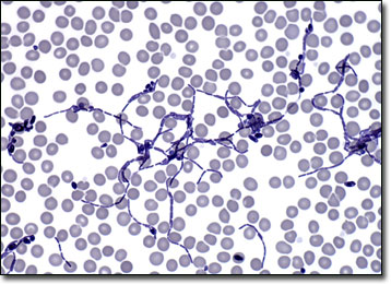
Despite the proliferation of antibacterial soaps and cleaners over the last decade, most bacteria are harmless or beneficial to humans. Indeed, the tiny organisms are significant contributors to the economies of many countries, aiding in the pickling, fermentation, and curing of a large number of products. Bacteria are also extremely valuable in the laboratory and have been utilized heavily in genetic research. Some bacteria are, however, pathogens that can cause disease in humans and other animals. These organisms are particularly adept at avoiding the body’s defense mechanisms once inside the bloodstream.
The vast majority of yeasts are members of the order Saccharomycetales in the class of Ascomycetes. Similar to bacteria, many varieties of the organisms can be quite valuable to humans. The single-celled asexual fungi, which reside in nature within soils and along plant tissues, are, for instance, widely utilized in the commercial manufacture of wine, beer, and breads. Yeast is also sold in small packets that are used in homes around the world to produce baked goods. A few varieties, such as Candida albicans, are, nevertheless, pathogenic and can cause various problematic conditions and infections.
BACK TO THE BRIGHTFIELD MICROSCOPY IMAGE GALLERY