
Brightfield Microscopy Digital Image Gallery
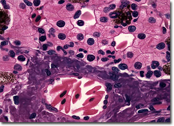
Salamander (Amphiuma) Liver
Salamanders may look like lizards, but they are not. The smooth-skinned animals, which typically inhabit moist areas near bodies of water, are unrelated to reptiles.
Salamanders are most prevalent in North America, but they may be found in other locales as well. In Japan, for instance, there exists a giant salamander that may grow more than five feet long. Most varieties of the animals are significantly smaller, however, typically exhibiting lengths of less than six inches. As adults, salamanders are usually terrestrial predators that primarily feed upon creatures smaller than themselves, such as worms and snails. Breeding usually takes place in water and the fertilized eggs grow into an aquatic larval form that undergoes the metamorphic process characteristic of amphibians.
Amphiuma is a genus of aquatic salamanders that have very small limbs and are sometimes misidentified as snakes or eels. Indeed, members of the genus, which may grow up to three feet long, are often better known by the name Congo eel or Congo snake. These salamanders spend most of their time burrowing through mud and debris searching for insects, mollusks, frogs, and other small prey, but they can also be dangerous to larger animals. Their teeth are quite sharp, their jaws are strong, and their bite can be brutal. Thus, anyone that comes into contact them should be wary.
BACK TO THE BRIGHTFIELD MICROSCOPY IMAGE GALLERY