Anatomy of the MIC-D Digital Microscope
Microscope Overview
The Olympus MIC-D digital microscope represents a unique design that incorporates a CMOS electronic digital imaging sensor as a substitute for the traditional eyepieces (commonly referred to as oculars) found in a majority of traditional microscopes. Coupled to an inverted illumination system that broadcasts light from above the specimen (similar to a tissue culture microscope), this digital microscope also features a translatable lamphouse and condenser unit that can be rotated over a 135-degree angle. Such versatility enables the operator to project light onto the specimen from an almost limitless combination of oblique or reflected illumination angles.
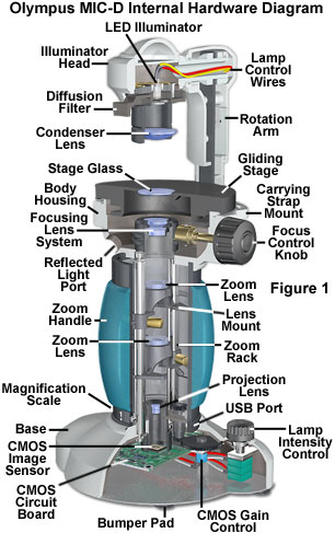
The important internal and external instrument configuration details of the Olympus MIC-D digital microscope are presented in Figure 1, which shows a cutaway diagram of the microscope including the electrical, mechanical, and optical components. Integral to the design is a Universal Serial Bus (USB) interface connection to a host computer that provides electrical current to power both the microscope illumination and a software interface to capture, edit, and catalog digital images acquired by the CMOS image sensor. Specimens are placed on the flat, circular stage and imaged with a zoom optical system controlled by a molded and cushioned rotation handle that forms part of the microscope body and contains a graduated reference magnification scale on the lower rim.
There are six basic mechanical control elements available to the operator for routine microscope configuration during specimen observation. The microscope can be powered on and off with a small potentiometer switch unit that is housed in the base of the microscope and also controls the lighting intensity. A single focus knob, positioned adjacent to the gliding stage, is utilized to bring the specimen into focus at any position throughout the entire magnification range. The stage is built with a gliding mechanism that allows the operator to rotate specimens, or translate them in virtually any direction. A diffusion filter (or screen) housed in the illumination head can be employed to alter the numerical aperture and distribution of light reaching the specimen, while the rotation arm can be translated to obliquely illuminate the specimen from a variety of angles.
The light source is a gallium-nitride based white light-emitting diode (LED) contained in the illumination head and powered by twin wire cables that snake through the body of the microscope to connect the illumination head and the electronic control circuitry housed in the base. Access to the LED is gained through a molded polymer Access Cover, which is threaded to fasten on top of the illumination head (see Figure 2). The lamp is mounted through a 5-millimeter aperture with two plastic-washered screws in a circular aluminum bracket within the illumination head, which is also machined from aluminum and internally anodized and dyed with a black pigment to absorb stray light.
The condenser lens element is seated in a cylindrical barrel housing that mounts to the underside of the lamp bracket. A rectangular notch in the upper portion of the condenser barrel serves as a guide way for the Diffusion Filter frame, which, like the access cover, is injection-molded from a high-impact acrylonitrile-butadiene-styrene (ABS) polymer. In the central portion of the diffusion filter frame are two 9-millimeter apertures, positioned adjacent to one another. One aperture holds the glass diffusion filter, while the other is opened to allow illumination from the lamp to pass through unhindered. The filter frame contains a 11.5 millimeter rectangular slot that acts as a guide for a pin limiting the forward and reverse motion of the frame in the illumination head. When the frame handle is pulled forward, the open aperture is placed in front of the lamp, and when the handle is pushed to the rear, light from the LED passes through the diffusion screen. The filter frame cannot be removed from the illumination head without disassembly of the entire unit.
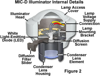
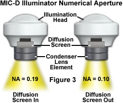
The condenser barrel contains a single bi-convex Lens Element (see Figure 2) having two unequal radii, with the larger-radius side facing the illumination source, and the side with greatest curvature facing the specimen. The numerical aperture and illumination intensity distribution is controlled by inserting or removing the diffusion filter from the microscope optical pathway. When the filter is inserted, the condenser has an effective numerical aperture of 0.19 (see Figure 3) to produce a broad cone of illumination necessary to match the objective numerical aperture values for brightfield observation throughout the zoom magnification range. Removing the diffusion filter limits the condenser numerical aperture to 0.10, but produces a more direct and intense level of illumination. This setting is optimal for highly oblique lighting, crossed polarized illumination, or reflected light directed through the substage port (illustrated in Figures 1 and 4).
The illumination head (Figures 2 and 3) is mounted onto the microscope rotation arm through a swivel mechanism that consists of an aluminum tube that seats into an opening drilled into the head. Lead wires that connect the LED lamp to the electronics in the microscope base pass through the central hollow section of the swivel tube. Surrounding the perimeter of the swivel tube at one end is a 1.5-millimeter channel that holds the four screws utilized to mount the swivel to the illumination head. The screws are positioned at 90-degree angles and anchor firmly into the swivel tube, which rotates over a 10-degree angle through an adjustable cam mechanism contained in the rotation arm. The illumination head range of motion on the swivel tube is controlled by two setscrews that limit the movement of the tube through flattened lands machined into a thick portion of the tube seat. The default motion range (10 degrees) is adequate for all off-axis illumination scenarios. Increasing this range can be accomplished by adjusting the set-screws, but will only produce a greater degree of chromatic and other less desirable off-axis aberrations from the condenser lens element when the illumination head is shifted too far.
The Rotation Arm (see Figure 1) consists of a flat metal bracket covered with a two-piece ABS polymer shroud having trim features that match the microscope design. The front section of the shroud is secured to the metal bracket with four Phillips head screws, and contains an opening for the illumination swivel arm. The shroud rear section is also fastened to the metal bracket with four similar screws, and covers the illumination wires that exit the swivel arm and travel the length of the bracket. These wires enter the microscope body through the main rotation arm attachment swivel joint. A small polymer wire connector is included about midpoint between the swivel tube and the main swivel joint to enable changeover of the LED light source.
The rotation arm is connected to the upper portion of the microscope body with a threaded, hollow flange bolt containing a flat brass washer and a thrust washer to ensure smooth motion. Wires from the LED lamp pass through the central part of the flange bolt and enter the microscope body through this port. A 4-millimeter diameter pin extends from the front of the rotation arm metal bracket and seats in a crescent-shaped radial groove or trench that is milled into the rear of the microscope body. The boundaries of the pin within the milled radial trench define the 135-degree range of motion for the rotation arm. When the pin is seated at one end of the trench, the rotation arm is parallel to the microscope optical axis for brightfield illumination, and when the pin is stopped at the other end, the illuminator is in the correct position to direct light through a lower port drilled into the microscope body (reflected illumination; see Figures 1 and 4).
The upper portion of the microscope body (Figure 4) is cast from aluminum as a single unit and machined to accept the stage, the rotation arm, the objective lens elements, the focus knob, and the rods that serve as guides for the zoom lens system. Cast into the upper body are two slotted openings that are utilized to mount a strap to carry the microscope. The outside surface of the unit has a rough texture and is painted, while the interior is covered with a blackened conversion coating that absorbs stray light.
| Interactive Tutorial | |||||||||||
|
|||||||||||
The gliding circular specimen Stage (98 millimeters in diameter; Figure 4) is machined from aluminum rod stock and the top is coated with a ceramic composite that is resistant to corrosion, detergents, and harsh chemicals. The underside of the stage is anodized and permeated with a black dye for a lower level of protection. A central 21-millimeter stage aperture contains a Glass Plate that is 1.05 millimeters thick and covered on both sides with a single-layer antireflection coating (illustrated in Figure 4). Light rays passing through the specimen travel through the glass plate and enter the objective front lens. The lower side of the plate also intercepts light passing to the specimen when the microscope is operated in reflected light mode.
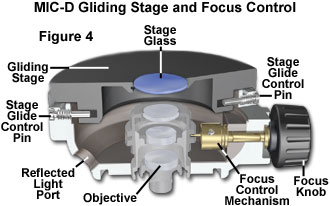
A U-shaped rim machined around the circumference of the lower stage serves as a seat for two guide pins (Glide Control Pins; Figure 4) that secure the stage to the microscope base. These pins restrict lateral stage motion, but enable the stage to be rotated in a gliding manner on top of the microscope base. The stage has a 7-millimeter range of motion in the x and y directions, but can orbit to any position within these boundaries. Beneath the stage glass is a set of threads designed to mount a polarizer for polarized light observations. The stage must be removed from the microscope body in order to add the polarizer, which can be accomplished by unseating the two threaded guide pins. When the stage is re-attached to the microscope base, check to ensure that the guide pins are properly installed in the stage rim.
Removing the stage exposes the movable objective front lens element and the Focus Control Mechanism, which are illustrated in Figure 4. Similar to the condenser lens, the objective front lens is bi-convex with surfaces having dissimilar radii. The surface having a larger radius (the "flatter" side of the lens) is almost flat and faces the specimen, while the surface with a smaller radius (greater curvature) faces the second objective lens element. This lens element is mounted in a machined black-anodized aluminum housing that has an extended annular ring with a groove cut into the center. A pin on the focus control mechanism fits into the groove and is used to translate the lens mount upward and downward when the focus knob is rotated.
The focus knob is mounted on a partially threaded shaft that is guided by a machined boss cast into the microscope upper body unit. A 10-millimeter length of brass rod is drilled and tapped to thread onto the focus knob shaft. On the end of the rod facing away from the knob is a small pin that fits into the lens mount groove. When the focus knob is rotated, the pin scribes an orbital path that translates the objective front lens to enable the operator to focus the specimen.
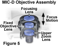
A second objective bi-concave lens element (see the MIC-D Objective Assembly illustrated in Figure 5) is mounted in a seat containing a 9-millimeter aperture machined into a boss cast in the center of the microscope optical axis. This lens element is fixed into position and serves to focus light from the front objective lens element into the zoom optical system. The aluminum mount holding the first objective lens glides over the cast aluminum boss (containing the second lens), which holds the upper objective assembly together.
Coupled to the two upper lens elements to form a working objective is the first lens in the zoom optical system, which serves as the final objective lens, and is seated in a counter-weighted translatable mount that glides up and down the body tube of the microscope as the magnification is varied (see Figure 6). A 9-millimeter diameter aperture diaphragm built into the mounting seat of this lens element acts as a primary conjugate plane for the illuminating (aperture) light flux passing through the microscope. The second lens of the zoom system performs the function of a relay lens to assist the projection lens (Figure 7) in forming a focused image of the specimen on the surface of the CMOS image sensor.
Zoom Magnification Range, Numerical Aperture, and Viewfield Size
|
||||||||||||||||||||||||||||||||||||||||||||||||||
Table 1
The optical zoom magnification range of the MIC-D microscope is presented in Table 1, along with the equivalent screen magnification indicated on the graduated scale located at the base of the Zoom Handle (see Figure 1). In addition, Table 1 lists the numerical apertures at corresponding objective magnifications, and the measured width of the Live Image screen on the host computer monitor. The objective magnification and numerical aperture ranges from 0.7x to 9.0x and 0.05 to 0.17, respectively, as the zoom lens system is racked from the lowest to the highest magnification range. Simultaneously, the viewfield shrinks from 6.9 millimeters at the lowest magnification to 630 micrometers at the highest (assuming a screen dot pitch of 0.3 millimeters). At any point within the zoom range, the magnification can be read directly from the graduated scale at the base of the zoom handle and the optical magnification calculated.
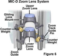
Each of the two zoom lens elements is seated in a specialized cast aluminum mount that enables them to be translated effortlessly on the two guideposts (Figure 6). The front (or upper) zoom lens mount has a larger diameter that enables it to snugly enter the objective boss that is cast into the microscope upper body unit. In addition, the recessed portion of this lens mount allows the lower zoom lens element to fit inside the first mount in order to bring the lenses together for close focusing at both ends of the zoom range. The mounts each contain offset brass counterweights that are designed to stabilize the movement of the lens mounts and eliminate image displacement from the center of the optical train when the zoom handle is rotated (and the lenses are moved up or down).
| Interactive Tutorial | |||||||||||
|
|||||||||||
On the end of the zoom lens mounts that contain the counterweights, a small pin is located that guides each lens mount in one of two grooved tracks inside the zoom handle body. The lens tracks trace a helical pattern within the zoom body and are positioned to produce accurate magnification factors and maintain focus throughout the zoom lens range. Two guideposts serve the dual purpose of maintaining the lens mounts at a stationary angle and securing the upper body unit to the microscope base. The posts are machined from stainless steel bar stock, then drilled and tapped at both ends for screws that attach them to the base and upper body unit. Countersunk holes in the upper body unit and microscope base anchor the guideposts in place. A third stainless steel post stabilizes the microscope and provides a foundation for the shielded wires that provide current for the LED light source. The wires are bound to this post with heat-shrink tubing to keep them from interfering with the moving zoom lens system.
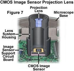
The recessed area of the lower zoom lens mount is designed to fit over the projection lens that directs the optical image to the surface of the CMOS image sensor (see Figure 7). The projection lens is a 2.4x magnification doublet housed in a cylindrical machined black-anodized aluminum mount with an outside diameter of 16 millimeters. When the zoom optical system is set to the lowest magnification position, the lower zoom lens element is placed directly above the projection lens. The lens mount is secured to the microscope base through a cutaway boss (cast into the base) with two mounting screws.
The lower end of the projection lens mount rests on the CMOS image sensor, which is installed on a circuit board and is aligned with the optical axis of the microscope. Light focused by the microscope optical system interacts with the CMOS photodiode array to generate electrical charge that is interpreted by the controlling electronics into an image. The circuit board containing the image sensor integrated circuit (see Figure 8) also contains the USB interface connector (and supporting electronics, including an integrated circuit) and a dynamic random access memory (DRAM) chip that buffers image data. The USB control integrated circuit transmits data from the CMOS image sensor to the host computer.
A second integrated circuit housed in the base of the microscope controls illumination intensity of the LED lamp. This circuit board is activated by the switch portion of the Lamp Intensity Control potentiometer, and the amount of current supplied to the lamp is controlled by the variable resistor. The switch also regulates power to the image capture board, described above. Both circuit boards are secured to the microscope base with screws, and are grounded to the aluminum casing with circular solder rings surrounding the mounting holes.
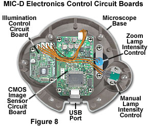
Lamp intensity is also controlled by the zoom handle through a series of planetary gears driven by a large circular gear molded into the circumference of the handle base. When the handle is rotated to adjust the microscope magnification, it simultaneously controls a potentiometer tied to the gear set and housed in the microscope base (the Zoom Lamp Intensity Control; see Figure 8). This potentiometer increases lamp intensity as the zoom system is racked to higher magnifications and reduces the intensity at lower magnifications. The electronics housed in the microscope base are covered with a flat aluminum plate (painted black on one side), which is secured to the base with small Phillips-head machine screws. Flexible synthetic polymer bumper feet cover the screw heads and provide a vibration-damper for the microscope.
In general, the Olympus MIC-D digital microscope is extremely well constructed, with carefully designed and fitted parts that require a considerable amount of pre-assembly preparation. Materials used in the construction appear to be very high in quality and are covered with paint, anodized with dyes, or treated with a conversion coating to absorb stray light and for protection. Although the MIC-D microscope is targeted at the entry-level student, the workmanship is reminiscent of that observed with research-level microscopes. Coupled with the unique design, which should set a standard for future microscopes in the coming years, the Olympus MIC-D affords a variety of contrast-enhancing techniques that are unparalleled in competing microscopes of this class.
Contributing Authors
Thomas J. Fellers and Michael W. Davidson - National High Magnetic Field Laboratory, 1800 East Paul Dirac Dr., The Florida State University, Tallahassee, Florida, 32310.
BACK TO MIC-D MICROSCOPE ANATOMY
BACK TO THE OLYMPUS MIC-D DIGITAL MICROSCOPE
Questions or comments? Send us an email.
© 1995-2025 by Michael W. Davidson and The Florida State University. All Rights Reserved. No images, graphics, software, scripts, or applets may be reproduced or used in any manner without permission from the copyright holders. Use of this website means you agree to all of the Legal Terms and Conditions set forth by the owners.
This website is maintained by our
Graphics & Web Programming Team
in collaboration with Optical Microscopy at the
National High Magnetic Field Laboratory.
Last Modification Tuesday, Sep 11, 2018 at 02:46 PM
Access Count Since September 17, 2002: 78317
Visit the website of our partner in introductory microscopy education:
|
|
