
Anatomy of the MIC-D Digital Microscope
Cutaway Microscope Diagram
The Olympus MIC-D digital microscope represents a unique design that incorporates a CMOS electronic digital imaging sensor as a substitute for the traditional eyepieces or oculars found in a majority of microscopes. Coupled to an inverted illumination system that broadcasts light from above the specimen (similar to a tissue culture microscope), this digital microscope also features a translatable lamphouse and condenser unit that can be rotated over a 135-degree angle. Such versatility allows the operator to project light onto the specimen from an almost limitless combination of oblique or reflected illumination angles.
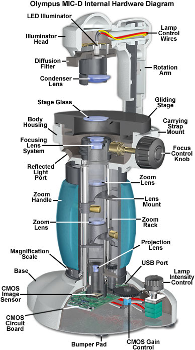
The important internal and external configuration details of the Olympus MIC-D digital microscope are presented in a large view in the figure above, which shows a cutaway diagram of the microscope including the electrical, mechanical, and optical components. Integral to the design is a Universal Serial Bus (USB) interface connection to a host computer that provides both electrical current to power the microscope, and a software interface to capture, edit, and catalog digital images acquired by the CMOS image sensor. Specimens are placed on the flat, circular stage and imaged with a zoom optical system controlled by a molded and cushioned rotation handle that forms part of the microscope body and contains a graduated reference magnification scale on the lower rim.
There are six basic mechanical control elements available to the operator for specimen observation. The microscope can be powered on and off with a small potentiometer switch unit that is housed in the base of the microscope and also controls the lighting intensity. A single focus knob, positioned adjacent to the gliding stage, is utilized to bring the specimen into focus at any position throughout the entire magnification range. The stage is built with a gliding mechanism that allows the operator to rotate specimens, or translate them in virtually any direction. A diffusion filter (or screen) housed in the illumination head can be employed to alter the numerical aperture and distribution of light reaching the specimen, while the rotation arm can be translated to obliquely illuminate the specimen from a variety of angles.
Contributing Authors
Thomas J. Fellers and Michael W. Davidson - National High Magnetic Field Laboratory, 1800 East Paul Dirac Dr., The Florida State University, Tallahassee, Florida, 32310.
BACK TO MIC-D MICROSCOPE ANATOMY
BACK TO THE OLYMPUS MIC-D DIGITAL MICROSCOPE
Questions or comments? Send us an email.
© 1995-2025 by Michael W. Davidson and The Florida State University. All Rights Reserved. No images, graphics, software, scripts, or applets may be reproduced or used in any manner without permission from the copyright holders. Use of this website means you agree to all of the Legal Terms and Conditions set forth by the owners.
This website is maintained by our
Graphics & Web Programming Team
in collaboration with Optical Microscopy at the
National High Magnetic Field Laboratory.
Last Modification Friday, Nov 13, 2015 at 02:19 PM
Access Count Since September 17, 2002: 30272
Visit the website of our partner in introductory microscopy education:
|
|