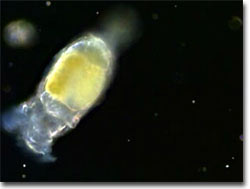
Philodina (Rotifera) Movies

Philodina Video No. 1 - Birth of a Philodina rotifer - a daughter emerges from her mother's body and swims off to start her new life; under darkfield illumination at a magnification of 100x with a playing time of 37.0 seconds. Choose a playback format that matches your connection speed: 28.8k (modem), 56.6k (modem), or T1/Cable/DSL, or download this video clip in MPEG format (6.43 MB).
Philodina Video No. 2 - Two Philodina rotifers feeding; under darkfield illumination at a magnification of 200x with a playing time of 16.5 seconds. Choose a playback format that matches your connection speed: 28.8k (modem), 56.6k (modem), or T1/Cable/DSL, or download this video clip in MPEG format (2.15 MB).
Philodina Video No. 3 - A translucent Philodina rotifer stretches out to feed, suddenly retracts, then stretches out in another area to feed; under darkfield illumination at a magnification of 200x with a playing time of 45.4 seconds. Choose a playback format that matches your connection speed: 28.8k (modem), 56.6k (modem), or T1/Cable/DSL, or download this video clip in MPEG format (14.1 MB).
Philodina Video No. 4 - With its food anchored in one spot, this Philodina rotifer stretches out to look for food; under phase contrast illumination at a magnification of 200x with a playing time of 26.3 seconds. Choose a playback format that matches your connection speed: 28.8k (modem), 56.6k (modem), or T1/Cable/DSL, or download this video clip in MPEG format (6.24 MB).
Philodina Video No. 5 - A Philodina rotifer extends and contracts its corona while feeding; under darkfield illumination at a magnification of 200x with a playing time of 17.9 seconds. Choose a playback format that matches your connection speed: 28.8k (modem), 56.6k (modem), or T1/Cable/DSL, or download this video clip in MPEG format (1.26 MB).
Philodina Video No. 6 - A Philodina rotifer jumps from one spot to another, making wormlike stretches in search of food; under phase contrast illumination at a magnification of 200x with a playing time of 31.3 seconds. Choose a playback format that matches your connection speed: 28.8k (modem), 56.6k (modem), or T1/Cable/DSL, or download this video clip in MPEG format (8.98 MB).
Philodina Video No. 7 - A Philodina rotifer explores for food, moving with wormlike stretches, then picking up its foot and jumping to another spot; under phase contrast illumination at a magnification of 200x with a playing time of 45.4 seconds. Choose a playback format that matches your connection speed: 28.8k (modem), 56.6k (modem), or T1/Cable/DSL, or download this video clip in MPEG format (10.7 MB).
Philodina Video No. 8 - Its foot anchored at one spot, a Philodina rotifer explores for food, moving with wormlike stretches, then picking up its foot and jumping to another spot; under phase contrast illumination at a magnification of 200x with a playing time of 33.6 seconds. Choose a playback format that matches your connection speed: 28.8k (modem), 56.6k (modem), or T1/Cable/DSL, or download this video clip in MPEG format (9.48 MB).
Philodina Video No. 9 - Two Philodina rotifers gobble up smaller microorganisms; under darkfield illumination at a magnification of 200x with a playing time of 44.9 seconds. Choose a playback format that matches your connection speed: 28.8k (modem), 56.6k (modem), or T1/Cable/DSL, or download this video clip in MPEG format (7.37 MB).
