Spike (M.I.) Walker
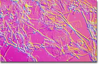
Candida albicans
English photomicrographer Spike (M.I.) Walker has been a consistent winner of the Nikon Small World competition for many years and has published many articles and a book about microscopy. Featured below is a differential interference contrast photomicrograph of the fungus Candida albicans.

|
The image above features Candida albicans in thrush infection of vaginal epithelium from a fresh scrape preparation. The objective was a planachromat 40x/0.65 NA coupled to a 1.4 numerical aperture achromatic differential interference contrast (DIC) condenser. Illumination was provided by a 12-volt, 100-watt tungsten-halide lamp. The microscope was a Zeiss Ultraphot IIIB with an automatic 35-millimeter photohead. Photographed with Fujichrome Velvia. (200x) |
Candida albicans is a dimorphic fungus that grows at 98.6 degrees Fahrenheit (37 degrees Celsius) and is the fungus that most often infects human beings. Normally, it lives on the mucosal membranes and the surface of the skin, causing little or no damage. In some circumstances, however, the same strains of C. albicans that grow as harmless commensals can become pathogenic, invading the mucosa and causing significant damage. This usually happens when a variety of predisposing factors cause the yeast population to multiply, escaping the normal competition from resident bacteria that keep the yeast population in check. When it invades the mucous membranes of the mouth or vagina, it produces a mild infection called thrush or candidiasis. The yeast cells sprout a hyphal outgrowth, which locally penetrates the mucosal membrane, causing irritation and shedding of the tissues. In patients whose resistance to infection is diminished, C. albicans can gain entry to the bloodstream and cause systemic candidosis.
Yeasts are fungi that grow as single cells, producing daughter cells either by budding (the budding yeasts) or by binary fission (the fission yeasts). They differ from most fungi, which grow as thread-like hyphae. However, some fungi alternate between a yeast phase and a hyphal phase, depending on environmental conditions. Such fungi are termed dimorphic (with two shapes) and they include several species, such as C. albicans, that cause disease of humans.
