
Spike (M.I.) Walker
Diatom (Actinoptychus)
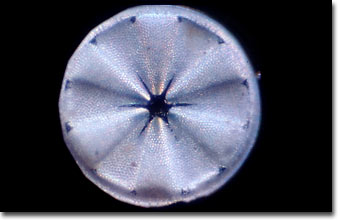
English photomicrographer Spike (M.I.) Walker has been a consistent winner of the Nikon Small World competition for many years and has published many articles and a book about microscopy. Featured below is a photomicrograph of the Diatom Actinoptychus.
|
The Actinoptychus specimen was photographed with a Metrimpex 3D condenser, a modified Abbe condenser of Hungarian manufacture, dating back to the early 1960s. The condenser has three interchangeable front lenses (brightfield, oil darkfield, and asymmetrical off-axis). A Zeiss microflash illumination system was used with a Reichert Zetopan Exacta camera body and the assistance of Rheinberg illumination filters. The film was Kodachrome 25. (50x) |
Diatoms have a silicified cell wall forms a pillbox-like shell (frustule) composed of overlapping halves that contain intricate and delicate markings useful in testing the resolving power of microscope lenses. The beautiful symmetry and exquisite design of diatom frustules have gained them the title "jewel of the sea."
Depending on the classification system used, diatoms are placed in either the phylum Bacillariophyta or in the class Bacillariophyceae in the phylum Chrysophyta (golden algae). Most diatoms are planktonic, but some are bottom dwellers or grow on other algae or plants. They are single-celled organisms, but some live in colonies. Diatoms are important components of phytoplankton, serving as primary sources of food for zooplankton in both marine and freshwater habitats. Diatomaceous earth, a substance composed of fossil diatoms, is used in filters, insulation, abrasives, paints, and varnishes.
These organisms usually reproduce asexually by cell division, the overlapping shell halves separate, and each secretes a smaller bottom half, decreasing in size with each division. Over a period of months there can be as much as a 60% decrease in size. Periodic spore formation serves to restore the diatom line to its original size.
Although diatoms are found in all of the Earth's aquatic environments, most species occur only in habitats with specific physical, chemical, and biological characteristics. Ecologists use this habitat specificity by collecting and analyzing individual species and community data to determine the quality or condition of aquatic habitats. Both long-term monitoring of specific lake and stream habitats and analysis of diatom remains (part of the sedimentary record of lakes) allow scientists to obtain a unique long-term historical perspective on these ecosystems.