Wim van Egmond
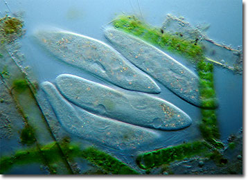
Paramecium
Paramecium is a genus of well-known protozoa whose members typically exhibit slipper-like shapes. Frequent visitors in classrooms and laboratories, an extensive amount of data regarding the microscopic organisms has been collected.

As ciliates, paramecia are covered with numerous, short hairlike projections called cilia. The beating of the cilia is what enables the microscopic organisms to locomote through their aquatic environments, as well as to procure their meals. Water currents created by the cilia sweep bacteria and other food particles into an individualís oral groove and gullet, where they begin the transformation into food vacuoles. Other characteristic structures found in paramecia include water-regulating contractile vacuoles, two different types of nuclei, and filamentous trichocysts, which may be involved in defense or extended for anchoring purposes during feeding.
In the strictest sense of the word, the only type of reproduction carried out by paramecia is the asexual process of binary fission. However, the protozoa are also capable of various sexual processes, such as conjugation, that involve the exchange of nucleic material, but do not directly result in an increase of numbers. During conjugation, two paramecia of compatible mating strains join together along their oral sides and a breakdown of their membranes facilitates the temporary formation of a shared cytoplasmic bridge. The micronuclei of both ciliates undergo several divisions and one from each travels across the bridge, where it fuses with a micronucleus of the opposing party. All other macro- and micronuclei then dissipate, leaving a single nucleus in both cells. Subsequently, the conjugating ciliates pull apart and their zygotic nuclei divide repeatedly to create both types of nuclei. This development is often immediately followed by the creation of new cells via fission.
BACK TO WIM VAN EGMOND GALLERY
