Wim van Egmond
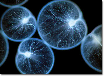
Seasparkle (Noctiluca) in Darkfield
Members of the genus Noctiluca are dinoflagellates, a class of primarily marine protozoa that possess two flagella. Dinoflagellates also characteristically feature a transverse groove, or girdle, that houses the circumferential flagellum and that exhibits a posterior extension called a sulcus that holds the longitudinal flagellum.

Noctiluca species are some of the largest single-celled organisms in the world. The most well known of the genus is Noctiluca scintillans, which may grow from 200 to 2000 microns in diameter. These dinoflagellates possess nearly spherical, balloon-like bodies that lack a pronounced girdle. The organisms do, however, retain two flagella, though they are highly modified, not always noticeable, and are not strong enough to readily facilitate locomotion. Rather, Noctiluca scintillans controls its vertical position by regulating its buoyancy, often floating just under the surface of the water.
Noctiluca scintillans is widely distributed throughout the world, occurring most often in coastal waters. Sometimes referred to as seasparkle, the organisms are often responsible for lighting up the sea, and sometimes the wet sand, at night. In large quantities, the dinoflagellates are bioluminescent, though individually they cannot produce perceptible light. The glittering phenomena produced by blooms is believed by some to be a defensive mechanism, acting as a deterrent to predators who want to avoid becoming bioluminescent themselves, an attribute that could make them more readily seen and caught by other organisms.
BACK TO WIM VAN EGMOND GALLERY
