
Wim van Egmond
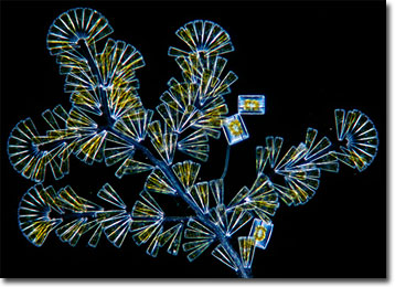
Licmophora Diatoms
Found in most every aquatic environment on the planet, the unicellular protists known as diatoms are important components of phytoplankton, serving as a primary food source for zooplankton. However, not all diatoms live a planktonic existence. Species may also be bottom dwellers, and some, like many of those belonging to the genus Licmophora, are epiphytes.
In addition to their substantial ecological importance, diatoms are noteworthy for their tremendous beauty. Sometimes referred to as the “jewels of the sea,” the microscopic organisms absorb silica from the environment and use it to secrete siliceous shells called frustules. These bivalve, glasslike shells, which are variously shaped and ornamented, have been of interest to microscopists for many years and are sometimes utilized to test the resolving power of microscopes. The picturesque frustules have also been used in a form of art, which was particularly popular during the Victorian period in England. By painstakingly arranging diatoms on permanent slides, creative microscopists fashioned stunning arrays and patterns that were to be enjoyed like miniature paintings. Indeed, many of these early pieces of diatom art can be found in modern museum collections.
The diatoms most people are familiar with tend to be circular, like Coscinodiscus, or distinctly pen- or cigar-shaped, like Navicula. Yet, even more beautiful forms may be found, such as the exquisite, fanlike shapes of Licmophora. Individuals within the genus grow from a common stalk, often attached to algae, rocks, or other substrates. When the organisms are particularly abundant on surfaces, they appear like short, yellow fuzz to the naked eye. It is only under the lens of a microscope that the splendor of the branching, colonial diatoms materializes.
BACK TO WIM VAN EGMOND GALLERY