
Wim van Egmond
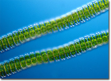
Desmidium swartzii
If variety is the spice of life, then desmids must exist in a constant state of delight. These predominantly unicellular green algae are well known for their tremendous diversity of cell shapes and sizes, which make it difficult for microscopists to find two of the organisms that are truly identical.
Beautifully symmetrical, desmids are typically divided into two mirror-image halves called semicells that are connected by an isthmus. Interestingly, the age of an individual desmid’s semicells is usually different. This seemingly strange phenomenon occurs because reproduction of the organisms is most often asexual, the centrally located nucleus dividing to create two daughter cells, each of which retains one of the original semicells. However, desmids are also capable of sexual processes, such as conjugation, which results in the formation of thick-walled zygotes that are able to withstand stressful environmental conditions.
Desmidium is a genus of filamentous desmids that inhabit freshwater bodies and are common components of the surface material frequently referred to as “pond scum.” The hairlike strands are composed of short cells with deep constrictions and do not feature the spiny projections that may appear among other related genera. A prominent member of the genus is Desmidium swartzii, which is characterized by colonies of triangular cells 11 to 22 micrometers long and 24 to 39 micrometers wide. Since the individuals are connected in such a way that they twist slightly, the colonies of the species generally exhibit a spiraling shape.
BACK TO WIM VAN EGMOND GALLERY