
Wim van Egmond
Coscinodiscus Diatoms
Described by German scientist Christian Ehrenberg in 1838 based on fossil specimens, Coscinodiscus is a genus of centric diatoms present in most bodies of water. Especially abundant in coastal areas, the free-living organisms are an important food source for microcrustaceans and a variety of other aquatic life forms.
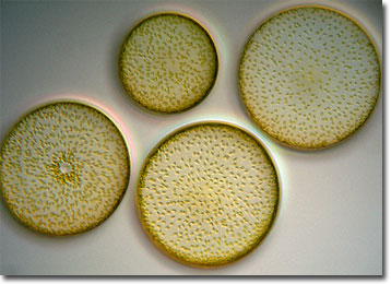
Taxonomically, diatoms are generally placed in either the algal phylum Bacillariophyta or in the class Bacillariophyceae of the protist phylum Chrysophyta. Tens of thousands of the chlorophyll-containing organisms have been identified in the fossil record, but only about 5,600 species are extant. The great abundance of diatom fossils is primarily due to the siliceous cell walls known as frustules that house them. Extremely durable, the frustules often display complex and intricate markings, a characteristic that has made them extremely popular with microscopists. In fact, the beautiful, symmetrical designs are sometimes even used to test the resolving power of microscopes.
Many diatoms are planktonic, but some species attach to substrates, such as plants or rocks, while others are bottom dwellers. The organisms range in size from approximately 2 micrometers to several millimeters, though only a few exhibit diameters greater than 200 micrometers. Typically, diatoms are divided into two basic categories: the radially symmetric Centrales, such as Coscinodiscus, and elongate, bilaterally symmetric Pennales. In addition to their disparity in shape and symmetry, the two orders of diatoms may differ in their ability to locomote. Often Centrales are considered to be nonmotile, while Pennales may move through aquatic environments with a gliding motion facilitated by mucilage secretions.
BACK TO WIM VAN EGMOND GALLERY