Loes Modderman
Decalcifier
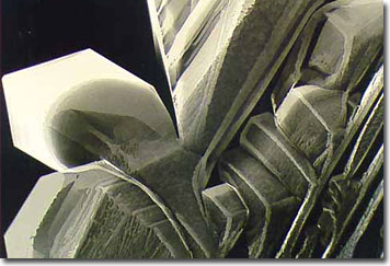
Decalcification is a technique for removing calcium from tissue specimens in order to permit medical and scientific observation, and this method is an important step in processing tissue and bone marrow biopsies. Preparing specimens for examination under microscopes is an important aspect of the decalcification process as well.

Calcium is a hard substance that must be removed before specimens are able to be cut into thin slices or sections. Without removal of calcium from bone or calcified tissues, specimen sections may not cut smoothly, and torn or ragged features may result. Cutting edges of fine instruments may be damaged as well.
Many decalcifying products contain additional chelating agents to remove minerals from tissue samples. A prime concern is that while removing calcium, fine cellular structures, and details of red blood cells, muscle tissue, or cancer cells may be lost. Buffers may be incorporated into solutions in order to prevent possible distortion and swelling of cells and diagnostic features. Solutions are also formulated so that specimens undergoing processing retain affinity for stains that are subsequently applied, so that details of cellular structures may be revealed.
BACK TO LOES MODDERMAN GALLERY
