Loes Modderman
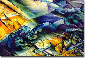
Calcium Carbonate
Calcium is a soft, silvery-white, metallic element first isolated by Sir Humphrey Davy in 1808. Occurring naturally in limestone, fluorite, and gypsum, this mineral is classified as an alkaline earth metal. As a primary constituent of bones, teeth, and shells, calcium phosphate is an essential mineral compound found in all living organisms.

Carbonate and phosphate salts, which are slightly soluble in water, are the primary constituents of bones. Acid-based decalcifying agents must be used to enhance the solubility of the metallic salts, and are designed to release calcium ions. Once mineral deposits are sufficiently softened, proper specimen sectioning can begin. Preparation of bones and other calcified tissue typically is achieved by soaking the sample for a required length of time in solutions that contain decalcifiers.
Many decalcifying products contain additional chelating agents to remove minerals from tissue samples. A prime concern is that while removing calcium, fine cellular structures, and details of red blood cells, muscle tissue, or cancer cells may be lost. Buffers may be incorporated into solutions in order to prevent possible distortion and swelling of cells and diagnostic features. Solutions are also formulated so that specimens undergoing processing retain affinity for stains that are subsequently applied, so that details of cellular structures may be revealed.
BACK TO LOES MODDERMAN GALLERY
