Fascinating Photos with
a Simple Microscope
Michael W. Davidson
National High Magnetic Field Laboratory (NHMFL)
and Center for Materials Research and Tech
The Florida State University
Tallahassee, USA

|
|
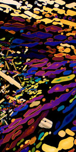
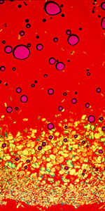
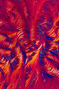
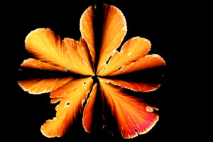
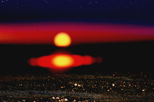
Photography through the microscope, or more commonly, photomicrography, has long been a useful tool for scientists. However, anyone who has access to a simple light microscope can produce highly color-saturated photographs, which display an exciting blend of art and science. Simple light microscopes with high-quality optics can be purchased for as little as $250, and adapters for most cameras are available for $25 to $75 (see the accompanying list of microscope manufacturers and distributors). Photographers who have access to state and government-surplus facilities may often find rather expensive surplus microscopes available at reasonable prices. Expensive high-powered microscope objectives (lenses) have a very narrow depth of field and are not really useful; therefore, a lower-cost 5x or 10x objective, which usually comes, as standard equipment on most microscopes, is all that is needed. All of the photomicrographs in this article were taken using a Nikon 4x or 10x objective. You can add polarizers to your microscope for just a few dollars, and the use of polarized light allows you to get beautiful photomicrographs of crystals made from common household chemicals.
|
|
GETTING STARTED
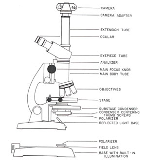
The first polarizer is inserted at the base of the substage condenser (shown in the microscope diagram on page 5) and can be held in place with tape. If the microscope has a built-in light source, the polarizer can be placed over the field lens. The second polarizer, commonly termed the analyzer, is placed inside the body of the microscope between the main-body tube and the eyepiece tube. There is usually a lens mount at the top of the body tube, and the analyzer can be placed directly on this mount. You will need a polarizer that is approximately 1-3cm in diameter because of the restricted area within the main body tube. This can be obtained by cutting a piece of polarized sheet plastic or buying a small polarizer from a science-supply distributor.
|
|
Next, rotate the polarizer until the viewfield becomes very dark. At this point, the polarization direction is perpendicular between the polarizer and the analyzer, and you have what is termed crossed polarizers. When purchasing polarizers, remember to select polarizers that are very close to a neutral gray in color, such as the threaded polarizers made for the front of a camera lens. Avoid polarizing materials that are green or amber in color.
|
|
It is very important to ensure that your microscope is aligned to produce even illumination across the viewfield. Information in microscope alignment is available in owner's manuals or in textbooks dealing with microscopy. An exceptionally good book, Photography Through the Microscope, is available from Kodak (Publication P-2), and can be purchased in many camera shops.
|
|
Most crystals are anisotropic and birefringment, which means that they will refract plane-polarized light emitted from the polarizer, and will bend it until it is visible through the analyzer when the polarizers are crossed.
To prepare crystals for examination in the microscope, you simply deposit a few granules of the crystal onto a microscope slide or a thin piece of 2 x 2-inch glass. Next, carefully place a microscope coverslip, easily obtained from a hobby shop or science-supply shop, over the crystals and heat slowly with a match or cigarette lighter until the crystals melt completely (a bunsen burner or alcohol lamp will do a very good job, if available). When melted, the crystals will flow underneath the coverslip and fill the entire volume between the coverslip and fill the entire volume between the coverslip and the microscope slide. Allow the slide to cool, and place it in a safe place for a few days to permit the melted chemical to slowly recrystallize. Very thick crystal samples will usually not transmit much light, and are not as colorful as thinner preparations. If you don't like the crystal formations after recrystallization, re-melt the sample and allow it to recrystallize a second time.
|
|
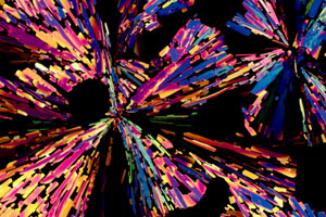
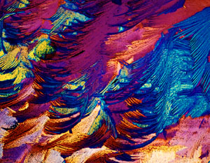
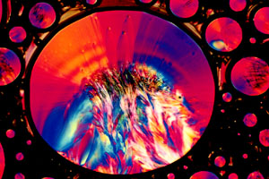
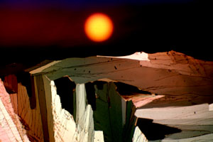
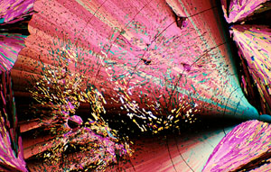
Another effective method of preparing crystals is to dissolve the chemical in a suitable solvent, like water, rubbing alcohol, or mineral spirits. This method is especially useful for chemicals in the salt family, like table salt, Epsom salts, Alka-Seltzer, and baking soda. Salts will usually decompose when heated, leaving a tarlike mess. However, when dissolved in water or alcohol, these salts will slowly recrystallize as the solvent evaporates to form colorful crystalline patterns. Many chemicals can form different types of crystals, depending on whether they are melt-recrystallized or recrystallized from solution. Some chemicals recrystallize almost immediately, while some take hours, days, or even weeks. The chemicals listed in the accompanying table are easily obtained, and will recrystallize in a reasonable amount of time to yield beautiful photomicrographs. Several examples of photomicrographs of household chemicals are illustrated.
|
|
CHOICE OF FILM
The majority of microscopes use a tungsten-halide bulb as a light source. These bulbs emit a very bright light with a wavelength spectrum centered in the 3200 K color-temperature range. There are a wide variety of films available that reproduce this color balance correctly.
|
|
Kodak's Ektachrome 50 and 160 are good examples of transparency films that are tungsten-balanced; however, I prefer the good contrast and color saturation provided by Fujichrome 64T. Photomicrographs are notoriously low in contrast, so I usually underexpose one or two f-stops and push E-6 processing one f-stop. This increases the contrast and color saturation, without a significant loss in density.

|
|
For Kodachrome lovers, Kodak makes a fine-grained ISO 40-speed, 3400 K-balanced Kodachrome, which can be corrected, for a tungsten-halide light source by the addition of a No. 82A filter between the light source and the first polarizer. If you are in a hurry, Polaroid's High Contrast Polachrome HCP (ISO 40) will produce very good results. With any film, it's wise to bracket the first roll to get a handle on exposure times.
|
|
Common chemicals suitable for recrystallization.
CHEMICAL COMMENTS
Alka-Seltzer - Best crystals from aqueous solution.
Aspartame (Nutra-sweet) - Good crystals from melt-recrystallization or aqueous solution.
Aspirin (acetylsalicylic acid) - Equally good crystals from melt-recrystallization or aqueous or ethanol solutions.
Acetaminophen (Tylenol) - Equally good crystals from melt-recrystallization or aqueous solution.
Epsom salts - Recrystallize from aqueous solution only.
Glucose and sucrose - Crystals quickly obtained (common sugars) by evaporation of solution in water. Very beautiful crystals after melt-crystallization.
Kodak Dektol paper developer - Best crystals after evaporation of working stock solution.
Kodak D-76 film developer - Best crystals after evaporation of working stock solution.
|
|
Microscope and accessory manufactures and distributors
|
OLYMPUS CORPORATION 4 Nevada Drive Lake Success, NY 11042 Telephone: 516-488-3880 |
CAROLINA BIOLOGICAL SUPPLY COMPANY 2700 York Road Burlington, NC Telephone: 800-334-5551 |
|
E. LEITZ, INC. 24 Link Drive Rockleigh, NJ 07647 Telephone: 201-767-1100 |
CARL ZEISS, INC. One Ziess Drive Thornwood, NY 10594 Telephone: 914-747-1800 |
|
NIKON INC. INSTRUMENT GROUP 623 Stewart Ave. Garden City, NY 11530 Telephone: 516-222-0200 |
AO SCIENTIFIC INSTRUMENTS P.O. Box 123 Buffalo, NY 14240 |
|
FISHER SCIENTIFIC 50 Fadem Road Springfield, NJ 07081 Telephone: 201-379-1400 |
EXCEL TECHNOLOGIES 90 Phoenix Avenue Enfield, CT 06082 |
|
EDMUND SCIENTIFIC CO. 101 E. Gloucester Pike Barrington, NJ 08007 Telephone: 609-573-6250 |
McCRONE ACCESSORIES AND COMPONENTS 850 Pasquinelli Dr. Westmont, IL 60559 Telephone: 312-887-7100 |
Michael W. Davidson is a Research Associate at the Institute or Molecular Biophysics at Florida State University.