Fabrication of Unusual Art Forms
With Multiple Exposure Color Photomicrography
Michael W. Davidson
Institute of Molecular Biophysics and
Center for Materials Research and Techology
Department of Physics
The Florida State University
Tallahassee, Florida 32306
Employing an assortment of classical and non-classical microscopy techniques I have succeeded in constructing a series of photomicrographs designed to resemble unusual and/or alien landscapes. I have termed these photomicrographs "microscapes" and have assembled a collection of approximately 1000 such photographs. By applying the techniques described in this article, the interested microscopist can create virtually an unlimited variety of microscapes.
INTRODUCTION
Photomicrography has long been employed as a medium for the recording and communication of scientific data in a broad spectrum of disciplines. Introduction of new transparency films with improved emulsions coupled to the progress made in optical coatings technology and computer-assisted exposure monitors has enabled the hotomicroscopist to acquire high quality photomicrographs which are deeply color-saturated and high in contrast. This has, in part, led to an explosion in the utilization of photomicrography throughout many new scientific areas previously unexplored by visible light microscopy. The growing interest in photomicrography as an art form is partially evidenced by the increasing number of photomicrograph contests held each year both on a domestic and international basis (1). Additionally, increased competition in the scientific trade journal and arbitrated scientific periodical market has resulted in a dramatic increase in the use of photomicrographs appearing on periodical front covers as an eye-catcher (2).
|
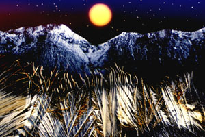
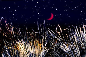
A multiple (5) exposure of ascorbic acid (the wheatfield in the foreground), liquid crystalline xanthin gum (the mountains), the field diaphram defocused with an orange the moon,), and polybenzyl-l-glutamate spherulites (the stars) with a blue filter. |
My interest in microscopy has led me to explore the possibilities of multiple exposure color photomicrography using crystals grown from a wide variety of chemicals, biochemicals, polymers, thin films, and biological macromolecules. The basic construction of these photomicrographs involves the classical microscopy techniques of brightfield and darkfield illumination, in addition to the now-standard methods of cross polarization, differential interference contrast, and Rheinberg illumination assisted by basic visible light color-filtering processes. This effort has resulted in the generation of a portfolio of images which I have termed microscapes. Microscapes consist of multiple exposures (so far from 2 to 9) fashioned on a single frame of 35mm transparency film. They are intended to resemble unusual and/or alien-like landscapes (see Figures 1-8) and are designed to have the highest contrast and color saturation currently available with commercial processing techniques. Other portfolios in my collection (3-5) contain images of various chemicals, biochemicals (sugars, vitamins, drugs, etc.), biological macromolecules, integrated circuits, memory devices, high-temperature superconductors, minerals, and thin films, however this discussion will center exclusively on the fabrication of microscapes.
MATERIALS AND METHODS
All photomicrographs were recorded on Fujichrome 64T 35mm transparency film operating at approximately ISO 64 when exposed to 3200° K color balanced light. Photomicrographs were exposed in either a Nikon Optiphot-pol polarizing transmitted light microscope equipped with a UFX-II exposure monitor or an Olympus BHSP reflected light microscope operating in the differential interference contrast or polarizing modes and operated by a PM-10AD exposure monitor. Exposures were usually made 1-3 f-steps below the recommended exposure on both the Nikon and Olympus monitors and Kodak E-6 processing was done in house. Slight modifications were made to the chemistry of the first and color developers to enhance contrast and color saturation. In addition, processing in the first developer was extended 25-30% over Kodak-recommended values to further enhance contrast and color saturation.
RESULTS AND DISCUSSION
The microscape collection of photomicrographs is centered around multiple exposure photography and utilizes a variety of both classical and non-classical microscopy techniques. Each micrograph was fabricated using both 4x (Nikon microscope or 5x on the Olympus microscope) and 10x objectives in a composite of multiple exposures on a single frame of 35mm film. Selective masking of previously exposed areas is critical to avoid undesirable overlap and washing out of successive exposures.
|
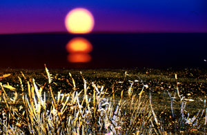
A multiple (5) exposure of a concentrated liquid crystalline DNA solution at the edge of a coverslip (the foreground intended to resemble plants at the edge of a lake), stretched polyethylene (the morning sky), the field diaphram defocused with a yellow filter in the lightpath (the rising sun), the field diaphram defocused with a diffraction grating in the lightpath (the sun's reflection), and liquid crystalline polybenzyl-l-glutamate spherulites (the stars). |
The first step in microscape construction is the exposure of a foreground which consists of a selected recrystallized chemical usually imaged with polarized light using the lower power objective and positioned so that only the bottom 30-50% of the film is exposed. Color transparency film is black when unexposed so the top half of the film remains black (unexposed) due to the crossed polarizers. A mask is cut (from a portion of black posterboard) to follow the profile of the first exposure and is placed directly on the field lens to selectively cover and prevent the first exposure from receiving any additional light.
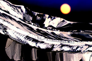
The second exposure can be an additional overlap on the first exposure (see Figure 3, for example), a crystalline formation resembling mountains (Figure 1), a seascape created with filters, or simply a simulated sky. Mountains can be simulated using a variety of recrystallized chemicals, although certain formations are more realistic than others. For instance, the snow-covered mountain effect illustrated in Figure 1 is obtained from a concentrated liquid crystalline solution of the polysaccharide xanthin in water. Another useful mountain background can be produced using crystals made from the organic buffer HEPES (3). Figure 5 is an example were the foreground and mountain were composed in a single crystal formation using HEPES.
|
A multiple (5) exposure of ascorbic acid slowly crystallized from aqueous ethanol and smeared across the microscope slide while drying (the sandy, rocky beach foreground), stretched polyethylene with a cyan filter (the water and morning sky), the field diaphram defocused with a yellow filter in the lightpath (the rising sun), and the field diaphram defocused with a fine-toothed comb inserted in the lightpath (the reflection of the sun). |
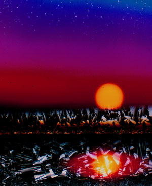
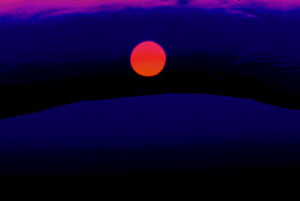
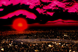
There are a variety of methods which can be applied to form diverse sky effects. After carefully cutting a mask to cover previous exposures, the microscope can be placed in the brightfield mode and a blue filter inserted into the lightpath (see Figures 1, 4, 5, and 7). A filter with very light intensity will work best because the color saturation of the sky can be controlled by reducing or increasing exposure times. Very short exposures yield a very dark blue sky while long exposures create a much lighter sky. It is very important here that the mask conforms to the topography of all previously exposed regions of the film. On exceedingly irregular boundaries like the mountains in Figure 1, a very short exposure time will result in a deep blue sky and avoid overlap of blue regions in the white mountains. A spectacular alternative to the blue filter technique involves cutting a 1 x 2 cm portion of a polyethylene sandwich baggie and stretching it to approximately 1 x 3 cm. This action tends to align the chain-like polyethylene molecules which enhances their birefringence. When viewed through cross polarizers, the aligned molecules combined with the thickness gradient created by the stretching induces the sheet of polyethylene to generate a prism-like effect. When the field is defocused, the sharp lines diffuse resulting in a yellow=>red=>blue morning sky effect as illustrated in Figures 2 and 3. A third effect which I take advantage of is the stormy bluish-purple cloudy sky outcome obtained by defocusing a bead of epoxy resin when imaged in polarized light with a 530 nm retardation plate positioned between the sample and the analyzer (Figure 8). Light diffracted by the mask accentuates the highlights producing an uncanny storm-like appearance. A blue filter can be added at the edge of the mask to make the effect more realistic (Figure 6).
|
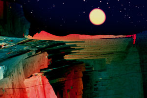
A multiple (4) exposure of sulfur crystals (the canyon-like foreground), the field diaphram defocused with a yellow filter in the lightpath (the moon), and liquid crystalline polybenzyl-l-glutamate spherulites (the stars) with a blue filter to simulate the sky. |
At this point, a sun or moon can be added to the micrograph. This is accomplished by closing the field diaphragm of the microscope until the desired sun or moon diameter is reached. Next, the image of the shutter leaves is defocused until they merge to form a complete circle. After placing the appropriate filter (usually orange, yellow, or red) in the lightpath, the image of the field diaphragm can be relocated to any position in the viewfield simply by adjusting the centering thumbscrews on the substage condenser. Placing a mask over the bottom portion of the image will create a rising sun effect as illustrated in Figures 2, 3, and 8. With a yellow or orange filter, the exposure time length determines the outcome of color distribution. Very short exposures produce images with a reddish perimeter and a saturated yellowish interior (Figures 1 and 2, for instance) while longer exposure times tend to wash out the image to yield more of a moon-like effect shown in Figure 4. Long exposure times when using a red filter will cause a yellowish center (not shown), while shorter times result in an even color effect (Figures 6 and 8).
|
A multiple (4) exposure of the biological buffer HEPES (the foreground and mountains in a single exposure), the field diaphram defocused with a yellow filter in the lightpath (the sun), and liquid crystalline polybenzyl-l-glutamate spherulites (the stars) with a blue filter to simulate the sky. |
To simulate the reflection of the sun or moon on water, two methods can be applied. After the diaphragm image is correctly placed, a diffraction grating can be inserted into the lightpath to spread out the image and shift the image colors to longer (redder) wavelengths (illustrated in Figure 2). I typically use Polachrome HC instant 35mm transparency film exposed to intense daylight because the manufacturing process for this film results in a series of very finely space lines which serve as an excellent diffraction grating when the film is greatly overexposed. Alternatively, a fine tooth comb inserted into the lightpath will produce the effect shown in Figure 3.
|
A multiple (2) exposure of the edge of an epoxy resin bead with a 530 nm retardation plate and blue filter (the mountain and sky) and the field diaphram defocused with a red filter (the sun). |
The new moon impression depicted in Figure 7 is produced simply by inserting the tip of a ball-point in the lightpath. A yellow filter used here instead of orange produces a more realistic appearance.
Generation of stars and/or clouds (not shown) is the final exposure in the series. After the field diaphragm exposure is complete, a sealed microscope slide containing a solution of small liquid crystalline spherulites of the polypeptide polybenzyl-l-glutamate is placed on the microscope stage. At low magnifications (10x), the spherulites appear as point-sources of light (Figure 2). By leaving the field diaphragm closed, an area on the slide can be located which is devoid of spherulites and this area is placed directly over the previously exposed diaphragm image area. This prevents stars from being imaged in the center of the moon or sun. After recentering and fully opening the field diaphragm, the exposure is made. Clouds are produced by defocusing colorless birefringent crystals of Cibachrome bleach. These crystals are usually imaged in the top-most portion of the viewfield and exposed for long times to wash out all color.
|
A multiple (4) exposure of melt-recrystallized ascorbic acid (the wheatfield),the field diaphram defocused with a yellow filter and a ball-point pin inserted into the lightpath (the moon), and liquid crystalline polybenzl-l-glutamate spherulites (the stars) with a blue filter to simulate the sky. |
Perhaps the most difficult shot is the beach scene (Figure 3) where the morning sky and ocean are created in a single exposure. This is accomplished by placing a blue filter over the mask at the field lens and defocusing the microscope until a sharp boundary occurs at the junction of the blue filter and the stretched polyethylene image. Correct positioning of the field diaphragm image to create a rising sun effect can be especially difficult when attempting to construct beach scenes.
|
A multiple (3) exposure of ascorbic acid slowly crystallized from aqueous ethanol and smeared across the microscope slide while drying (the sandy, rocky foreground), an epoxy resin bead with a 530 nm retardation plate and blue filter (the sky) and the field diaphram defocused with a red filter(the rising sun). |
It is not necessary to follow this outline exactly in order to obtain nicely formed microscapes. For instance, when I observe an unusually promising crystalline formation I usually compose enough images to fill several 24-exposure magazines of film. In this case, I proceed through the sequence as outlined above for the first series of exposures (to produce the intended microscape), then reverse the procedure constructing another very similar microscape thus minimizing the number of substage condenser centering operations. Because the sun or moon is not relocated during recording of the two successive microscapes, almost identical copies are obtained. This is especially beneficial if the resulting arrangement of exposures is optimal. With the proper equipment on hand for the assembly of microscapes, the interested microscopist is limited only by the boundaries of his own imagination.
REFERENCES
1. The following contests are held each year to promote photomicrography and provide an expanded base of marketing images for the sponsors.
NIKON SMALL WORLD CONTEST, held by Nikon Inc. Instrument Group, 623 Stewart Avenue, Garden City, New York 11530.
POLAROID INTERNATIONAL INSTANT PHOTOMICROGRAPHY CONTEST, held by the Polaroid Corporation, 575 Technology Square, 9P, Cambridge, Massachusetts 02139.
AMERICAN SOCIETY OF CLINICAL PATHOLOGY MEDICAL PHOTOGRAPHY COMPETITION, sponsored by Nikon, Inc., held by the American Society of Clinical Pathologists, 2100 West Harrison Street, Chicago, Illinois 60612.
OLYMPUS VISIONAGE INTERNATIONAL PHOTOGRAPHY CONTEST, held by the Olympus Optical Co., Ltd., San-Ei Bldg., 22-2 Nishi-Shinjuku, 1-Chome, Sinjuku-Ku, Toyko 163-19, Japan.
BETHESDA RESEARCH LABORATORIES' Life Sciences Photography Contest, held by the Bethesda Research Laboratories and Life Technologies, Inc., P.O. Box 6009 Gaithersburg, Maryland 20877.
INDUSTRIAL PHOTOGRAPHY PHOTO ANNUAL, held by Industrial Photography Magazine. PTN publishing Co., 210 Crossways Park Dr., Woodbury, New York 11797.
Royal Microscopical society micrograph competition, 3738 St. Clements, Oxford OX4 1AJ, England.
2. The following is a partial list of periodicals which have recently adopted photographs and photomicrographs on the front covers:
| ACTA ANATOMIC |
| AMERICAN CERAMIC SOCIETY BULLETIN |
| AMERICAN FAMILY PHYSICIAN |
| the AMERICAN JOURNAL OF PATHOLOGY |
| AMERICAN JOURNAL OF PHYSIOLOGY |
| the ANATOMICAL RECORD |
| ANGEWANTE CHEMIE |
| ANNALS OF APPLIED BIOLOGY |
| AUSTRALIAN JOURNAL OF PLANT PHYSIOLOGY |
| BIOESSAYS |
| BIOLOGIST |
| BIOLOGY OF THE CELL |
| BIOPHYSICAL JOURNAL |
| BIOSCIENCE |
| BIOTECHNOLOGY PROGRESS |
| CHIMIA |
| CELL |
| CELL BIOLOGY |
| CELL DIFFERENTIATION AND DEVELOPMENT |
| CELL STRUCTURE AND FUNCTION |
| CHEMISTRY AND INDUSTRY |
| CHEMISTRY IN BRITAIN |
| CHEMTECH |
| CHROMOSOMA |
| CONSERVATION BIOLOGY |
| CRITICAL REVIEWS IN MICROBIOLOGY |
| CRITICAL REVIEWS IN TOXICOLOGY |
| DEVELOPMENT |
| DEVELOPMENTAL BIOLOGY |
| DEVELOPMENTAL NEUROSCIENCE |
| DIFFERENTIATION, ONTOGENY AND NEOPLASIA |
| ENDEAVOUR |
| EUROPEAN JOURNAL OF BIOCHEMISTRY |
| EUROPEAN JOURNAL OF PARASITOLOGY |
| EXPERIMENTAL CELL RESEARCH |
| EXPERIMENTAL NEUROLOGY |
| the FASEB JOURNAL |
| GENES AND DEVELOPMENT |
| GEOSCIENCE CANADA |
| INTERNATIONAL ARCHIVES OF ALLERGY AND |
| APPLIED IMMUNITY |
| INTERNATIONAL JOURNAL OF LIPID RESEARCH |
| INTERVIROLOGY |
| INVERTEBRATE REPRODUCTION AND DEVELOPMENT |
| JOURNAL OF THE AMERICAN CERAMIC SOCIETY |
| JOURNAL OF BIOLOGICAL EDUCATION |
| JOURNAL OF BIOMOLECULAR STRUCTURE AND DYNAMICS |
| JOURNAL OF CELL BIOLOGY |
| JOURNAL OF CELL SCIENCE |
| JOURNAL OF CHEMICAL EDUCATION |
| JOURNAL OF CLINICAL INVESTIGATION |
| JOURNAL OF COMPARATIVE NEUROLOGY |
| JOURNAL OF GEOLOGICAL EDUCATION |
| JOURNAL OF HEREDITY |
| JOURNAL OF MATERIALS SCIENCE |
| JOURNAL OF MOLECULAR BIOLOGY |
| JOURNAL OF NEUROSCIENCE |
| JOURNAL OF NEUROCYTOLOGY |
| JOURNAL OF NEUROPATHOLOGY AND EXPERIMENTAL NEUROLOGY |
| JOURNAL OF NEUROSURGERY |
| JOURNAL OF PHYSICS D. APPLIED PHYSICS |
| JOURNAL OF PROTEIN CHEMISTRY |
| JOURNAL OF THE TEXTILE INSTITUTE |
| the LARYNGOSCOPE |
| MATERIALS ENGINEERING |
| the PLANT CELL |
| PHOTONICS SPECTRA |
| PROTEINS, STRUCTURE, FUNCTION & GENETICS |
| PROTOPLASMA |
| RENAL PHYSIOLOGY AND BIOCHEMISTRY |
| SYMBIOSIS |
| SVENSK BOTANISK TIDSKRIFT |
| TRENDS IN BIOCHEMICAL SCIENCES |
| TRENDS IN GENETICS |
| TRENDS IN PHARMACEUTICAL SCIENCES |
| TRENDS IN NEUROSCIENCES |
| WELDING JOURNAL |
3. Article entitled "My Dreams Are of Landscapes Made with Vitamin C" in FOTO CREATIV #3, May/June 1990, a West German photography magazine. A photomicrograph with a HEPES-generated mountain is featured on the front cover and a four page illustrated interview with the author appears on pages 72-75.
4. Michael W. Davidson and Randolph L. Rill, "Photomicrography. Common Ground For Science and Art", MICROSCOPY and Analysis May 1989, issue #11 pages 7-12.
5. Michael W. Davidson, "Fantastic Photography With A Simple Light Microscope", PHOTOgraphic Magazine in press (1990).
