
Twentieth Century Microscopes
Early in the twentieth century, microscope manufacturers began marketing parfocal objectives, allowing the image to remain in focus when the microscopist changed magnification by exchanged objectives on the rotating nosepiece. In 1924, Zeiss introduced a LeChatelier-style metallograph with infinity-corrected optics, but this method of correction would not see widespread application for another 60 years.
Shortly before World War II, Zeiss created several prototype phase contrast microscopes based on optical principles advanced by Frits Zernike. Several years later the same microscopes were modified to produce the first time-lapse cinemicrography of cell division photographed with phase contrast optics. This contrast-enhancing technique did not become universally recognized until the 1950s and is still a method of choice for many cell biologists today.
Physicist Georges Nomarski introduced improvements in Wollaston prism design for another powerful contrast-generating microscopy theory in 1955. This technique is commonly referred to as Nomarski interference or differential interference contrast (DIC) microscopy and, along with phase contrast, has allowed scientists to explore many new arenas in biology using living cells or unstained tissues. In 1975, Robert Hoffman introduced another method of increasing contrast in living material by taking advantage of phase gradients near cell membranes. This technique is now termed Hoffman Modulation Contrast, and is available as optional equipment on most modern microscopes.
The majority of microscopes manufactured around the world had fixed mechanical tube lengths (ranging from 160 to 210 millimeters) until the late 1980s, when manufacturers largely migrated to infinity-corrected optics. The past decade has witnessed an enormous growth in the application of optical microscopy for micron and sub-micron level investigations in a wide variety of disciplines. Rapid development of new fluorescent labels has accelerated the expansion of fluorescence microscopy in laboratory applications and research. Advances in digital imaging and analysis have also enabled microscopists to acquire quantitative measurements quickly and efficiently on specimens ranging from photosensitive caged compounds and synthetic ceramic superconductors to real-time fluorescence microscopy of living cells in their natural environment. Optical microscopy, with help of digital video, can also be used to image very thin optical sections, and confocal optical systems are now in operation at most major research institutions. At the close of the century, optical microscopy enjoyed a widespread renaissance in the scientific community, and late in 1999, computer giant Intel teamed with toy guru Mattel to produce perhaps the most remarkable microscope ever invented, the $100 Intel Play QX3 Computer Microscope.
Download our Museum of Microscopy screen saver for Windows.
Twentieth Century Bausch & Lomb Microscopes - When the twentieth century rolled around, Bausch & Lomb was the leading producer of microscopes in the United States, and the third largest manufacturer in the world (after Leitz and Zeiss). The StereoZoom series of microscopes is perhaps the most famous from the Bausch & Lomb inventory, although the company produced a number of high quality microscopes over a period exceeding 100 years. In the late 1980s, Bausch & Lomb entered into negotiations with Leica to sell the microscope division, which ultimately became incorporated into the Ernst Leitz name.
James Swift & Son Compound Dissecting Microscope - Designed to produce an upright image for minute dissections, this microscope was built around 1903 by James Swift & Son of 313 High Holborn in London.
Italian Heliostat/Colimator Solar Microscope - Described by museum curators as a heliostat, this instrument is lacking a mechanism to follow the sun's movement. The microscope is probably of Italian origin and made sometime in the early 1900s.
W. Watson and Sons Compound Monocular Microscope - An intricately detailed microscope with a large number of individual adjustments, this enameled and brass microscope was made in 1904.
Andrew Ross Compound Monocular Microscope - Originally termed the Standard No. 1 when introduced in the early 1900s, this brass microscope embodied the latest features of the period.
Upright Compound Student Microscope - This basic iron and nickel upright compound microscope was useful to students and amateur microscopists alike during the early 1900s. Part of the Billings collection at the Walter Reed Army Hospital in Washington, DC, the unsigned monocular instrument is not attributed to any craftsman, firm, or country.
Spencer Compound Monocular Microscope - Designated as Model 26B, this compound microscope was introduced by the Spencer Lens Company in 1904.
Watson & Sons Compound Monocular Microscope - Housed in a wooden-framed glass box, this continental-style horizontal compound microscope was modeled after nineteenth century French and German horizontal box microscopes.
Nikon's First Microscope - This simple brass and enamel compound monocular microscope is the forefather of a grand line of microscopes that rank among today's most sophisticated optical instruments.
Carl Zeiss Brain Section Microscope - Modeled after the popular Zeiss Stand ID design, this microscope was produced sometime around 1907.
Leitz Photomicrographic Apparatus of 1910 - This unique marriage of early twentieth century microscopy and photographic technology was very advanced for the period.
Powell and Lealand Iron Stand Microscope - Originally described as the Last Powell Iron Stand by Edward Milles Nelson, a famous British microscopist and former President of the Royal Microscopical Society, the Powell and Lealand iron stand monocular, compound microscope was commissioned in 1911 for Augustus Alfred Cornwallis Eliot Merlin.
Charles Baker Compound Microscope - British microscope designs were quite similar to each other at the beginning of the twentieth century, and this example is typical of microscopes of the period.
Monocular Compound Dissecting Microscope - Designed by Professor A. Gandolfi Hornyold, this simple monocular microscope was intended for studies of freshwater pond life.
Simple Compound Tripod Microscope - Inscribed "Junior: Made in Germany", this cute compound microscope is supported by a tripod and is equipped with three interchangeable objectives.
Winkel-Zeiss 2 Dissecting Microscope - In 1911, Carl Zeiss and the Rudolf Winkel microscope firm of G�ttingen, Germany formed a partnership and separate corporation known as R. Winkel GmbH, which thrived and grew to 360 employees before the post-war crash of 1945. The Winkel-Zeiss 2 dissecting microscope featured below is a product of their joint venture and was designed to meet the needs of beginning biology students in the 1920s.
Ernst Leitz Dual Objective Binocular Microscope - Used widely for dissection and manipulation of small objects, this 1920's stereomicroscope was equipped with dual objectives.
Zeiss Laboratory Microscope - A workhorse for the ages, this microscope is still in service in many laboratories around the world.
Spencer Compound Binocular Microscope - The unusual stage design on this microscope provides a convenient fine-focus mechanism to aid the operator in examining specimens.
Leitz Monocular Microscope - Similar in design and execution to the 1930s-model Zeiss, this Leitz microscope was serious competition during the period.
Newman's Compound Microscope - An ingenious design made from a hodgepodge of discarded microscope parts.
Edinburgh Student Microscope - Designated the model H, by builders W. Watson & Sons, this elaborate microscope was made for medical students and demonstrates advanced design motifs of the period.
Baker Microslide Projector - Designed to project magnified images of microscopical specimens, the Charles Baker microslide projector features a small, horizontal, compound microscope mounted with an electric illumination system on a kidney-shaped iron base.
Carl Zeiss Ultraphot II Microscope System - Perhaps one of the most beautiful microscopes ever designed, this Zeiss microscope was a world leader in workmanship and quality during the 1950s.
Leitz Inverted "Chemists Microscope" - Designed in the 1950s and continued until the mid 1970s, this microscope was originally intended to examine chemical reactions, but instead found widespread use as a tissue culture microscope.
StabiFocal Compound Microscope - An unusual microscope design signed StabiFocal Locquin Paris-France that, while innovative, did not succeed in the microscope market.
Siemens Elmiskop IA Transmission Electron Microscope - This all-purpose electron microscope addressed ease-of-use problems by being one of the first "user-friendly" instruments of the period.
Carl Zeiss Universal Microscope - Designed with both transmitted and reflected light illumination globes, this classic microscope is still serving microscopists in laboratories around the world today.
Nikon Model M Inverted Microscope - Built in the early 1970s, this black-enameled inverted microscope was designed for both biological (diascopic) and metallurgical (episcopic) use.
McArthur's Portable Compound Microscope - Deviating in design and construction from other instruments of the period, this rectangular microscope was given a design award in 1970.
Olympus Vanox Microscope - This early 1970s microscope was the flagship of the Olympus America lineup.
Nikon Diaphot Inverted Microscope - An advanced mid-1980s inverted tissue culture microscope that was readily adapted for enhanced contrast, photomicrography, and micromanipulation techniques.
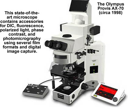
The Olympus Provis AX-70 - This high-end microscope from Olympus is considered to be the latest state-of-the-art in microscope design and construction.