
Zeiss Laboratory Microscope
Carl Zeiss has been a respected name in optical engineering and technology for over 150 years, and many of their microscopes are among the finest ever made. The microscope illustrated below is a standard laboratory microscope, introduced by Zeiss in the 1930s, that has proven to be a workhorse for both routine lab work and advanced research investigations as evidenced by the fact that many are still in service today.
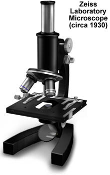
The microscope stand is constructed with a sturdy cast iron frame that is polished and coated with a hard coat of black enamel. The extra weight of this frame stabilizes the microscope and reduces the de-focusing effects of vibration. Three objectives can be positioned in the revolving nosepiece, and the sample is viewed through a Huygenian eyepiece positioned in a single monocular body tube. The substage condenser usually corrects for a low to medium degree of chromatic aberration and the concave mirror below the stage is designed to optimize illumination of the specimen. Focusing is achieved through a rack and pinion gear set enabled with a knurled knob where the body tube attaches to the stand. The stage is an X-Y mechanical stage with translation afforded in both directions by rack work mechanisms. A spring-loaded clip is used to secure the specimen to the mechanical stage holder. This microscope can be adapted for photomicrography by addition of the appropriate adapters to the eyepiece socket.
BACK TO TWENTIETH CENTURY MICROSCOPES