Microscopy Primer
Light and Color
Microscope Basics
Special Techniques
Digital Imaging
Confocal Microscopy
Live-Cell Imaging
Photomicrography
Microscopy Museum
Virtual Microscopy
Fluorescence
Web Resources
License Info
Image Use
Custom Photos
Partners
Site Info
Contact Us
Publications
Home

The Galleries:

Photo Gallery
Silicon Zoo
Pharmaceuticals
Chip Shots
Phytochemicals
DNA Gallery
Microscapes
Vitamins
Amino Acids
Birthstones
Religion Collection
Pesticides
BeerShots
Cocktail Collection
Screen Savers
Win Wallpaper
Mac Wallpaper
Movie Gallery

Carl Zeiss Brain Section Microscope

This microscope is signed "Carl Zeiss, Jena, No. 33362", and was designated the brain section microscope when introduced sometime around 1902. The model featured below was redrawn from a photograph of the original microscope, which is part of the Billings microscope collection at Walter Reed Army Hospital in Washington DC.

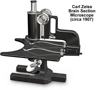
With a sturdy horseshoe base, this microscope was derived from the popular Zeiss Stand ID design. The large stage is almost 10 inches square and is incurved at the front with two very large spring clips to secure specimens. Such a large stage requires an abnormal projection of the microscope limb, which positions the body tube in approximately the center of the stage. A single ocular and objective occupy each end of the body tube, which is focused by a rack and pinion mechanism controlled by a pair of knurled brass knobs. Beneath the stage, attached to the stand, is a substage condenser and plane mirror. Accessories include a triple revolving nosepiece and spare objectives.

BACK TO TWENTIETH CENTURY MICROSCOPES

Questions or comments? Send us an email.
© 1998-2022 by Michael W. Davidson and The Florida State University. All Rights Reserved. No images, graphics, scripts, or applets may be reproduced or used in any manner without permission from the copyright holders. Use of this website means you agree to all of the Legal Terms and Conditions set forth by the owners.
This website is maintained by our
Graphics & Web Programming Team
in collaboration with Optical Microscopy at the
National High Magnetic Field Laboratory.
Last modification: Friday, Nov 13, 2015 at 02:19 PM
Access Count Since April 12, 2000: 28856
For more information on microscope manufacturers,
use the buttons below to navigate to their websites: