Microscopy Primer
Light and Color
Microscope Basics
Special Techniques
Digital Imaging
Confocal Microscopy
Live-Cell Imaging
Photomicrography
Microscopy Museum
Virtual Microscopy
Fluorescence
Web Resources
License Info
Image Use
Custom Photos
Partners
Site Info
Contact Us
Publications
Home

The Galleries:

Photo Gallery
Silicon Zoo
Pharmaceuticals
Chip Shots
Phytochemicals
DNA Gallery
Microscapes
Vitamins
Amino Acids
Birthstones
Religion Collection
Pesticides
BeerShots
Cocktail Collection
Screen Savers
Win Wallpaper
Mac Wallpaper
Movie Gallery

Edinburgh Student Microscope

W. Watson & Sons termed this microscope model H, when it was first introduced for medical students in May, 1934. The illustration below has been redrawn from photographs of the original microscope, which is featured in Gerard Turner's excellent book Collecting Microscopes, a volume in Christie's International Collectors Series of books on antiques.

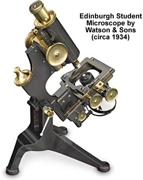
The base is a sturdy tripod that supports the limb (and the rest of the microscope) through a locking swivel joint. The body tube contains a single interchangeable brass objective at one end, and a brass eyepiece at the other. Focus is achieved through a brass rack and pinion mechanism that moves with entire body with respect to the limb. The elaborate stage is equipped for mechanical translation of the specimen in both the X and Y directions, and the substage condenser is centerable and has a height adjustment. Beneath the condenser is a double mirror unit that is attached to the lower limb through a gimbal. This is one of the more intricate designs made for students, but demonstrates the degree of microscope sophistication in the mid-1930s.

BACK TO TWENTIETH CENTURY MICROSCOPES

Questions or comments? Send us an email.
© 1998-2022 by Michael W. Davidson and The Florida State University. All Rights Reserved. No images, graphics, scripts, or applets may be reproduced or used in any manner without permission from the copyright holders. Use of this website means you agree to all of the Legal Terms and Conditions set forth by the owners.
This website is maintained by our
Graphics & Web Programming Team
in collaboration with Optical Microscopy at the
National High Magnetic Field Laboratory.
Last modification: Friday, Nov 13, 2015 at 02:19 PM
Access Count Since January 9, 2000: 45802
For more information on microscope manufacturers,
use the buttons below to navigate to their websites: