Interactive Tutorial
Image-Forming and Aperture Light Pathways
The Olympus MIC-D digital microscope is equipped with a zoom lens system that operates in conjunction with the objective lens elements to form an image of the specimen on the surface of the CMOS image sensor. This interactive tutorial explores both the image-forming and aperture light ray pathways through the microscope zoom optical body and how they are affected by changing the position of individual lens elements.
The tutorial initializes with a cutaway diagram of the MIC-D digital microscope, which reveals the internal zoom lens elements and the optical ray trace pathways through the microscope. To operate the tutorial, use the Magnification slider to translate the zoom lens system between the lowest (0.7x) and highest (9.0x) magnification values. As the slider is moved from left to right, the approximate magnification appears above the slider bar. Two radio buttons, entitled Image-Forming Light Rays and Aperture (Pupil) Light Rays, determine the light ray paths that are presented within the zoom body. When the Image-Forming Light Rays radio button is active (the default condition), imaging ray traces are shown in the tutorial. Activating the Aperture (Pupil) Light Rays radio button will show these ray paths.
In order to simplify and reduce the size of the illumination system, the MIC-D digital microscope lacks the condenser and field diaphragm apertures necessary to establish Köhler illumination. Therefore, the microscope must rely on an alternative configuration that does not require these aperture diaphragms to achieve uniform illumination of the specimen in order to fill the objective aperture. Olympus engineers chose the critical (often termed source focused or Nelsonian) method of illumination for the MIC-D, which was first developed by British microscopist Edward Nelson using optical principals advanced by Ernst Abbe. Critical illumination was successfully employed by microscopists from the latter part of the nineteenth century until well into the twentieth century. Even today, there are still some advocates of critical illumination who continue to use the method with microscopes requiring external light sources, or cheaper student microscopes where photomicrography or digital imaging is not an issue.

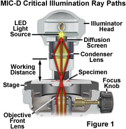
Critical illumination in the MIC-D digital microscope relies on using the condenser lens in the illumination head to produce a focused, real image of the light source (the LED) in the plane of the specimen to achieve a uniform illumination distribution over the entire viewfield (illustrated in Figure 1). Homogeneity of the light source is the important aspect when considering this method of illumination. Prior to the invention of electric lamps, microscopists were limited in their choice of suitable sources for microscope illumination. During daylight hours, they could point their microscopes (or substage reflector mirrors) towards the sky and use the clouds as a crude diffusion screen to spread illumination evenly across the entire field of view. Indoor and night work forced early microscopists to rely on artificial sources of illumination, such as oil lamps. The flame produced by a burning lamp is fairly even and consistent, but other sources, including frosted enlarger bulbs, opal bulbs, ribbon filaments, and light emitting diodes can also be utilized for critical illumination.
In order to align a microscope for critical illumination, light emitted from the source must be focused by the substage condenser so that a real image of the source is produced in the specimen plane at the microscope slide (Figure 1). The illumination numerical aperture must match that of the objective to ensure that the specimen assumes the optical properties of self-luminosity. In practice, it is often difficult (or impossible) to find focus in the central portion of diffuse light sources (such as a flame or the epoxy jacket of a light emitting diode), so the "edge" of the source is usually focused with subsequent readjustment of the substage mirror so that the image of the central portion of the light source fills the field of view. In the MIC-D digital microscope, the LED light source is fixed into position with respect to the specimen, and is pre-adjusted at the factory. The amount of light entering the microscope is controlled by adjusting the intensity of the LED through the potentiometer on the base of the microscope or by sliding the diffusion screen into or out of the light path. Enough light must enter the microscope to completely fill the rear focal plane of the objective, and this can be controlled by increasing voltage using the LED intensity potentiometer after inserting the diffusion screen into the optical pathway.

Focusing the light source in the specimen plane is precarious and can often yield a grainy, uneven, or speckled background unless the diffusion screen is inserted into the light path. On compound microscopes, the same effect can be produced by slightly defocusing the substage condenser to produce a more uniform background, but in these instruments, critical illumination has largely been supplanted by the far more efficient Köhler method of microscope illumination. In brightfield microscopy (as well as all other forms), efficient sample illumination is very dependent upon proper alignment of all the optical components of the microscope, including the illumination source. Uneven illumination can have a serious impact on the quality of digital images, causing hot spots, vignetting, color fringes, poor contrast, and a variety of other undesirable effects. Because the MIC-D digital microscope is equipped with a pre-centered lamp (that does not allow for adjustment), the only variables in illumination are the presence or absence of the diffusion screen and the voltage supplied to the light emitting diode.
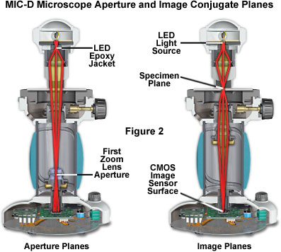
In critical illumination, the light source forms an image on the specimen, which resides in an image (or field) plane. There are three primary image planes in the MIC-D digital microscope (see Figure 2), the LED light source, the specimen plane, and the surface plane of the CMOS image sensor. Each of these planes forms a focused conjugate image of the specimen. The first lens of the zoom optical system is actually the final objective lens, and is seated in a counter-weighted translatable mount that glides up and down the body tube of the microscope as the magnification is varied. An aperture diaphragm built into the mounting seat of this lens element acts as a primary conjugate plane for the illuminating (aperture) light flux passing through the microscope. The second lens of the zoom system performs the function of a relay lens to assist the projection lens in forming a focused image of the specimen on the surface of the CMOS image sensor. A second conjugate aperture plane is situated between the LED and the diffusion screen (Figure 2). Thus, the MIC-D microscope has three principal image conjugate planes and two aperture (or illumination) conjugate planes. The location of these planes differs from those found in conventional microscopes that operate under the principles of Köhler illumination, but they work in combination to perform similar functions.
Contributing Authors
Matthew J. Parry-Hill and Michael W. Davidson - National High Magnetic Field Laboratory, 1800 East Paul Dirac Dr., The Florida State University, Tallahassee, Florida, 32310.
BACK TO MIC-D MICROSCOPE ANATOMY
BACK TO THE OLYMPUS MIC-D DIGITAL MICROSCOPE
Questions or comments? Send us an email.
© 1995-2025 by Michael W. Davidson and The Florida State University. All Rights Reserved. No images, graphics, software, scripts, or applets may be reproduced or used in any manner without permission from the copyright holders. Use of this website means you agree to all of the Legal Terms and Conditions set forth by the owners.
This website is maintained by our
Graphics & Web Programming Team
in collaboration with Optical Microscopy at the
National High Magnetic Field Laboratory.
Last Modification Tuesday, Sep 11, 2018 at 02:51 PM
Access Count Since September 17, 2002: 21642
Visit the website of our partner in introductory microscopy education:
|
|
